Clinical TrailsNovartis RWE: Pluvicto Delivers 13.5-Month PFS in ARPI-Pre-Treated mCRPC Novartis RWE: Pluvicto Delivers 13.5-Month PFS in ARPI-Pre-Treated mCRPC Read Post »
Health Tidings Policy & AcquisitionsAstellas and Vir Biotechnology Unite to Advance VIR-5500, a Next-Gen T-Cell... Astellas and Vir Biotechnology Unite to Advance VIR-5500, a Next-Gen T-Cell... Read Post »
Health TidingsTeva/Medincell Advance: SOLARIS-Backed TEV-‘749 NDA Accepted by FDA Teva/Medincell Advance: SOLARIS-Backed TEV-‘749 NDA Accepted by FDA Read Post »
Health TidingsNovo Nordisk Slashes List Prices of Wegovy, Ozempic, and Rybelsus by Up to ... Novo Nordisk Slashes List Prices of Wegovy, Ozempic, and Rybelsus by Up to ... Read Post »
Clinical TrailsNovo Nordisk-Backed Triple Agonist UBT251 Delivers Strong Phase 2 Results i... Novo Nordisk-Backed Triple Agonist UBT251 Delivers Strong Phase 2 Results i... Read Post »
Health Tidings Policy & AcquisitionsOrganon Licenses MIUDELLA®: New Hormone-Free Copper IUD Option Organon Licenses MIUDELLA®: New Hormone-Free Copper IUD Option Read Post »
Health TidingsFlexible SC Amivantamab Regimens Now Authorized EU-Wide for EGFR-Mutant Lun... Flexible SC Amivantamab Regimens Now Authorized EU-Wide for EGFR-Mutant Lun... Read Post »
Clinical TrailsMoonLake’s SLK Shows Strong Results in Phase 2 Trial for Axial Spondy... MoonLake’s SLK Shows Strong Results in Phase 2 Trial for Axial Spondy... Read Post »
Clinical TrailsGossamer Bio’s PROSERA Trial: Narrow Miss on 6MWD, Strong in Advanced... Gossamer Bio’s PROSERA Trial: Narrow Miss on 6MWD, Strong in Advanced... Read Post »
Clinical TrailsCagriSema Delivers 23% Weight Loss in REDEFINE 4: Strong Efficacy, Just Shy... CagriSema Delivers 23% Weight Loss in REDEFINE 4: Strong Efficacy, Just Shy... Read Post »
Health TidingsUltragenyx Scores FDA Priority Review for DTX401 in Rare Metabolic Disorder Ultragenyx Scores FDA Priority Review for DTX401 in Rare Metabolic Disorder Read Post »
New Drug ApprovalFDA Approves Zepbound 4-Dose KwikPen for Obesity Care FDA Approves Zepbound 4-Dose KwikPen for Obesity Care Read Post »
Clinical Trails Policy & AcquisitionsGilead Snaps Up Arcellx, Eyes Anito-cel Approval in Multiple Myeloma Gilead Snaps Up Arcellx, Eyes Anito-cel Approval in Multiple Myeloma Read Post »
Health TidingsEMA Validates ENHERTU® for Post-Neoadjuvant HER2+ Breast Cancer EMA Validates ENHERTU® for Post-Neoadjuvant HER2+ Breast Cancer Read Post »
Clinical TrailsFrom Week 44 to 140: New TREMFYA® Evidence Reinforces Long-Term UC Control... From Week 44 to 140: New TREMFYA® Evidence Reinforces Long-Term UC Control... Read Post »
Health TidingsSetback for Corcept as Court Backs Teva in Korlym Generic Fight Setback for Corcept as Court Backs Teva in Korlym Generic Fight Read Post »
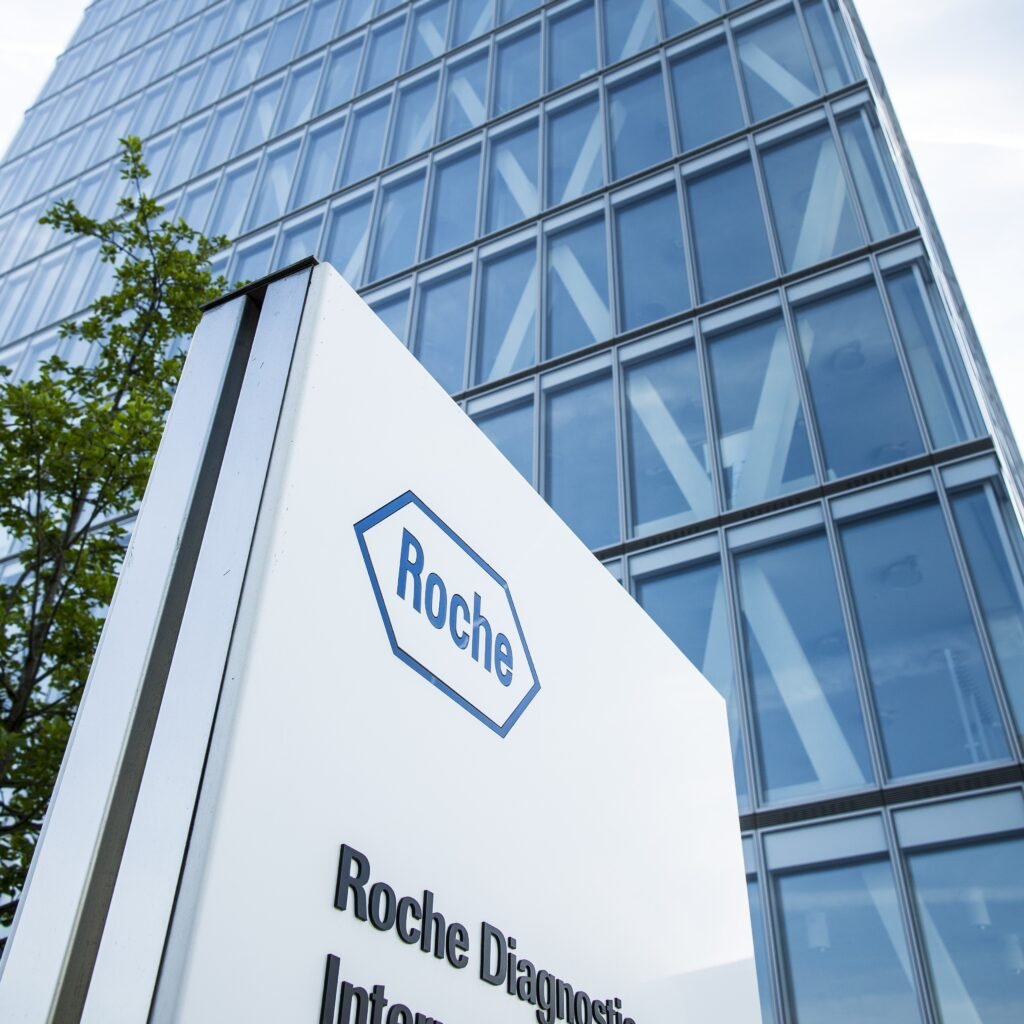
Health TidingsRoche’s Giredestrant NDA Accepted by FDA for ESR1-Mutated Breast Canc... Roche’s Giredestrant NDA Accepted by FDA for ESR1-Mutated Breast Canc... Read Post »
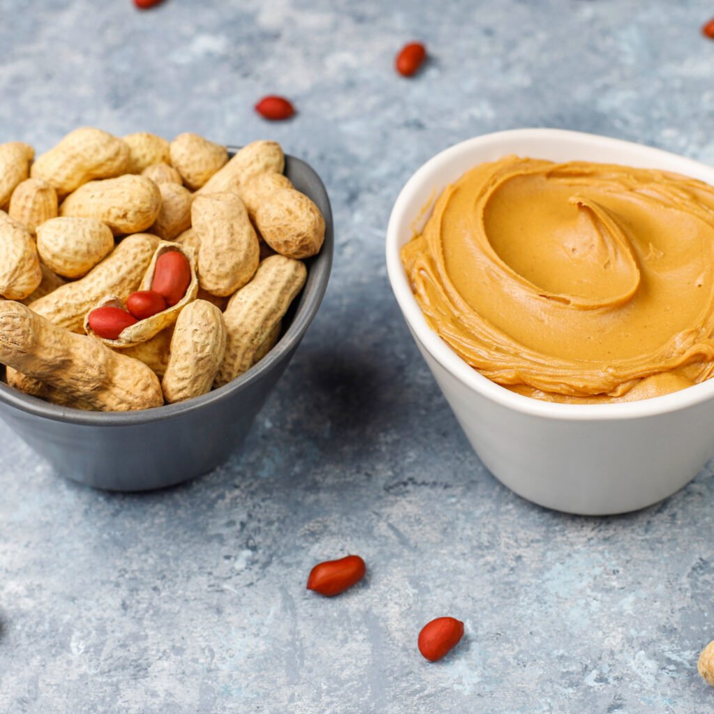
FoodPeanut Butter: A Balanced Look at Its Health Benefits, Risks, and Everyday ... Peanut Butter: A Balanced Look at Its Health Benefits, Risks, and Everyday ... Read Post »
Drugs Safety AlertNew GBS Safety Information Added to GSK’s SHINGRIX Label New GBS Safety Information Added to GSK’s SHINGRIX Label Read Post »
Health TidingsSavara’s MOLBREEVI BLA Accepted by FDA with Priority Review for Rare ... Savara’s MOLBREEVI BLA Accepted by FDA with Priority Review for Rare ... Read Post »
Health TidingsOne High-Quality Trial, Not Two: what the FDA changed and what it means One High-Quality Trial, Not Two: what the FDA changed and what it means Read Post »
Health TidingsOutlook Therapeutics Partners with Mediconsult for Exclusive LYTENAVA™ Di... Outlook Therapeutics Partners with Mediconsult for Exclusive LYTENAVA™ Di... Read Post »
New Drug ApprovalFDA Approves BYSANTI™: A New Ally in Bipolar I and Schizophrenia Treatmen... FDA Approves BYSANTI™: A New Ally in Bipolar I and Schizophrenia Treatmen... Read Post »
New Drug ApprovalChugai report Anti-CD20 Monoclonal Antibody Rituxan® Approved for AIHA Chugai report Anti-CD20 Monoclonal Antibody Rituxan® Approved for AIHA Read Post »
Clinical TrailsTakeda KEPLER Trial: Promising Vedolizumab Results for Pediatric Ulcerative... Takeda KEPLER Trial: Promising Vedolizumab Results for Pediatric Ulcerative... Read Post »
Health TidingsChugai Launches ELEVIDYS in Japan for Duchenne Muscular Dystrophy Chugai Launches ELEVIDYS in Japan for Duchenne Muscular Dystrophy Read Post »
Health TidingsRYBREVANT FASPRO™ Shines in First-Line HNSCC with 56% Response Rate RYBREVANT FASPRO™ Shines in First-Line HNSCC with 56% Response Rate Read Post »
Health TidingsOmvoh Sets New IBD Standard: 3-Year Data + Lilly’s Combination Pipeli... Omvoh Sets New IBD Standard: 3-Year Data + Lilly’s Combination Pipeli... Read Post »
New Drug ApprovalAbbVie’s VENCLEXTA Taps AZ’s Acalabrutinib for FDA’s Firs... AbbVie’s VENCLEXTA Taps AZ’s Acalabrutinib for FDA’s Firs... Read Post »
New Drug ApprovalEuropean Commission Grants Conditional Approval for ANKTIVA® in BCG-Unresp... European Commission Grants Conditional Approval for ANKTIVA® in BCG-Unresp... Read Post »
Health TidingsBoehringer Ingelheim Teams Up with NIPER Raebareli for Pharma Research Boos... Boehringer Ingelheim Teams Up with NIPER Raebareli for Pharma Research Boos... Read Post »
Clinical TrailsViiV Healthcare’s Cabenuva Demonstrates Superior Efficacy in LATITUDE... ViiV Healthcare’s Cabenuva Demonstrates Superior Efficacy in LATITUDE... Read Post »
Health TidingsFDA Grants Priority Review to Garetosmab for Rare Bone Disorder FOP FDA Grants Priority Review to Garetosmab for Rare Bone Disorder FOP Read Post »
Clinical TrailsMerck’s ENFLONSIA Shows Promising Safety in Second RSV Season for Hig... Merck’s ENFLONSIA Shows Promising Safety in Second RSV Season for Hig... Read Post »
Health Tidings Policy & AcquisitionsNovartis Licenses UNP’s Macrocyclic Peptides in $1.8B CVD Collaborati... Novartis Licenses UNP’s Macrocyclic Peptides in $1.8B CVD Collaborati... Read Post »
Clinical TrailsPfizer Advances BRAFTOVI in Colorectal Cancer with New BREAKWATER Results Pfizer Advances BRAFTOVI in Colorectal Cancer with New BREAKWATER Results Read Post »
Clinical TrailsNovartis’ Remibrutinib Shows Promise in Chronic Inducible Urticaria Novartis’ Remibrutinib Shows Promise in Chronic Inducible Urticaria Read Post »
Health TidingsJanux Therapeutics Dosed First Patient in JANX011 Study Janux Therapeutics Dosed First Patient in JANX011 Study Read Post »
Health TidingsRTF Resolved: Moderna’s Vaccine mRNA-1010 Advances to FDA Review RTF Resolved: Moderna’s Vaccine mRNA-1010 Advances to FDA Review Read Post »
Health TidingsFDA Accepts BMS’s NDA for Iberdomide in RRMM FDA Accepts BMS’s NDA for Iberdomide in RRMM Read Post »
Health TidingsEli Lilly’s TOGETHER-PsO Trial: Taltz Plus Zepbound Delivers Superior... Eli Lilly’s TOGETHER-PsO Trial: Taltz Plus Zepbound Delivers Superior... Read Post »
Health TidingsCompass Pathways Achieves Breakthrough in Phase 3 Trial for Psilocybin-Base... Compass Pathways Achieves Breakthrough in Phase 3 Trial for Psilocybin-Base... Read Post »
New Drug ApprovalFDA Approves Simplified Monthly Dosing for RYBREVANT FASPRO™ in EGFR-Muta... FDA Approves Simplified Monthly Dosing for RYBREVANT FASPRO™ in EGFR-Muta... Read Post »
Clinical TrailsViiV Healthcare Highlights Lotivibart and Cabotegravir LA Findings at CROI ... ViiV Healthcare Highlights Lotivibart and Cabotegravir LA Findings at CROI ... Read Post »
Health TidingsGSK RSV Vaccine Shows 75.6% Effectiveness Against Hospitalizations in Real-... GSK RSV Vaccine Shows 75.6% Effectiveness Against Hospitalizations in Real-... Read Post »
Clinical TrailsOcular Therapeutix’s AXPAXLI Achieves Superiority in Wet AMD Phase 3 ... Ocular Therapeutix’s AXPAXLI Achieves Superiority in Wet AMD Phase 3 ... Read Post »
New Drug ApprovalGSK’s Exdensur Gains EU Approval for Severe Asthma and CRSwNP GSK’s Exdensur Gains EU Approval for Severe Asthma and CRSwNP Read Post »
Clinical TrailsSanofi and Teva Report Durable Duvakitug Efficacy at 44 Weeks in UC/CD: REL... Sanofi and Teva Report Durable Duvakitug Efficacy at 44 Weeks in UC/CD: REL... Read Post »
Health Tidings Policy & AcquisitionsKidswell Bio and Treehill Launch Kidswell USA to Advance Cell Therapy for C... Kidswell Bio and Treehill Launch Kidswell USA to Advance Cell Therapy for C... Read Post »