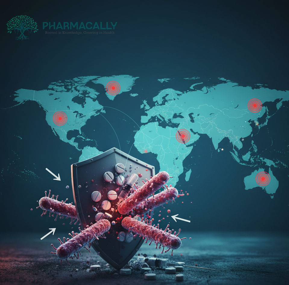
ResearchWhat the WHO 2025 Antibiotic Resistance Report Warns: The World’s Fight a... What the WHO 2025 Antibiotic Resistance Report Warns: The World’s Fight a... Read Post »
ResearchFSSAI Bans Misleading Use of ORS Label on Fruit Juices: Dr. Sivaranjani San... FSSAI Bans Misleading Use of ORS Label on Fruit Juices: Dr. Sivaranjani San... Read Post »
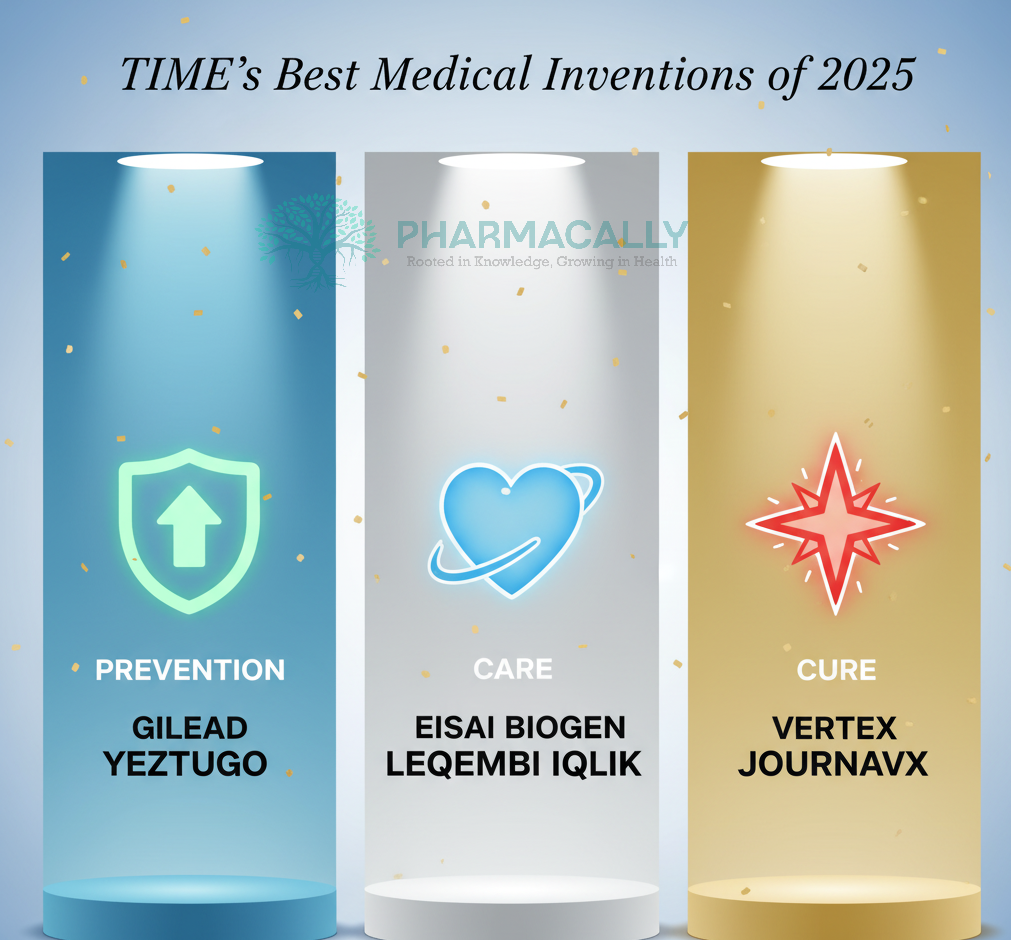
ResearchVertex’s Journavx, Eisai Biogen’s Leqembi Iqlik, and Gilead’s Yeztugo... Vertex’s Journavx, Eisai Biogen’s Leqembi Iqlik, and Gilead’s Yeztugo... Read Post »
ResearchJAMA Study Finds Asthma Inhalers Have a Carbon Footprint Equal to 530,000 C... JAMA Study Finds Asthma Inhalers Have a Carbon Footprint Equal to 530,000 C... Read Post »
ResearchWhat Is Wockhardt’s New Drug Zidebactam? A Groundbreaking Beta-Lactam Enh... What Is Wockhardt’s New Drug Zidebactam? A Groundbreaking Beta-Lactam Enh... Read Post »
Research2025 Nobel Prize in Physiology or Medicine: Pioneering Discoveries in Immun... 2025 Nobel Prize in Physiology or Medicine: Pioneering Discoveries in Immun... Read Post »
ResearchRethinking Lithium: A Potential Therapeutic Avenue in Alzheimer’s disease Rethinking Lithium: A Potential Therapeutic Avenue in Alzheimer’s disease Read Post »
Drugs Safety Alert ResearchGlobal Acetaminophen and Autism Concerns: Controversy Explained Global Acetaminophen and Autism Concerns: Controversy Explained Read Post »
Disease & Drugs ResearchCan Mouth Bacteria Trigger Heart Attacks? New Research Says Yes Can Mouth Bacteria Trigger Heart Attacks? New Research Says Yes Read Post »
Health Tidings ResearchIndian State of Kerala Reports Brain-Eating Amoeba Cases: A rare infection ... Indian State of Kerala Reports Brain-Eating Amoeba Cases: A rare infection ... Read Post »
Health Tidings ResearchFDA to Take Stricter Action on Misleading Drug Ads, Says Commissioner FDA to Take Stricter Action on Misleading Drug Ads, Says Commissioner Read Post »
ResearchGlobal Cholera Situation in 2024: A Worsening Public Health Crisis-A WHO re... Global Cholera Situation in 2024: A Worsening Public Health Crisis-A WHO re... Read Post »
ResearchThe Halifax Declaration: Protecting Health, Dignity, and Human Rights of Re... The Halifax Declaration: Protecting Health, Dignity, and Human Rights of Re... Read Post »
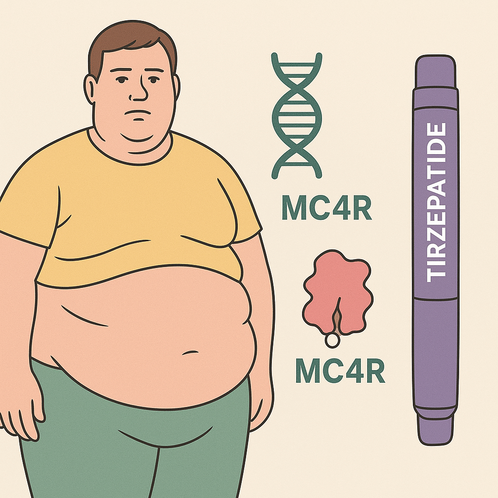
ResearchTirzepatide Shows Weight Loss Benefits Even in People with Genetic Obesity ... Tirzepatide Shows Weight Loss Benefits Even in People with Genetic Obesity ... Read Post »
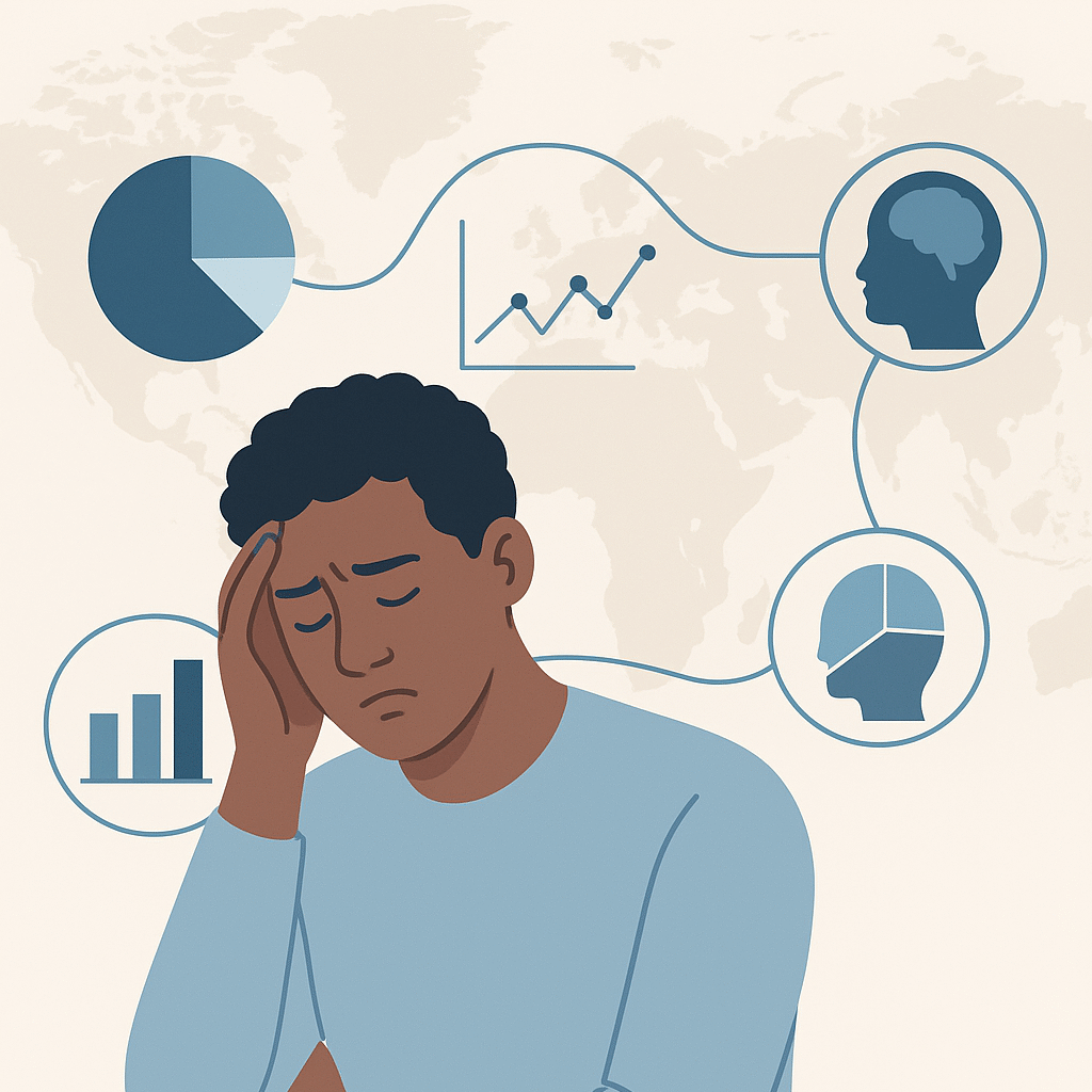
ResearchGlobal Mental Health in 2025: Evidence, Gaps, and the Way Forward-Analysis ... Global Mental Health in 2025: Evidence, Gaps, and the Way Forward-Analysis ... Read Post »
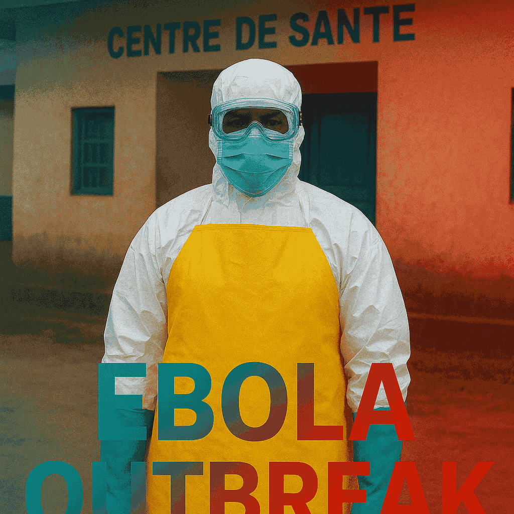
Health Tidings ResearchNew Ebola outbreak in Kasai province of DR Congo-WHO Confirmed New Ebola outbreak in Kasai province of DR Congo-WHO Confirmed Read Post »
ResearchEvery Life Matters: WHO Suicide Global Health Report 2021 Every Life Matters: WHO Suicide Global Health Report 2021 Read Post »
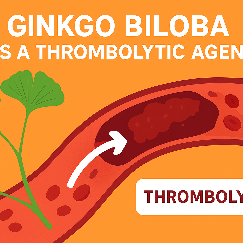
Research“Ginkgo Biloba as a Thrombolytic Agent: Can It Replace Aspirin in Long-Te... “Ginkgo Biloba as a Thrombolytic Agent: Can It Replace Aspirin in Long-Te... Read Post »
ResearchShingles Vaccine May Protect the Heart for Up to Eight Years, Landmark Sout... Shingles Vaccine May Protect the Heart for Up to Eight Years, Landmark Sout... Read Post »
ResearchTaiwan Study: Heatwaves Age People Like Smoking or Alcohol Taiwan Study: Heatwaves Age People Like Smoking or Alcohol Read Post »
Research“Repurposing Fenbendazole in Oncology: Hype vs. Hope in the Context o... “Repurposing Fenbendazole in Oncology: Hype vs. Hope in the Context o... Read Post »
ResearchWearable Health Technology: Turning Pharmacovigilance from Reactive to Proa... Wearable Health Technology: Turning Pharmacovigilance from Reactive to Proa... Read Post »
ResearchThe Hidden Pain Pathway: Paracetamol (Acetaminophen) Metabolite AM404 Block... The Hidden Pain Pathway: Paracetamol (Acetaminophen) Metabolite AM404 Block... Read Post »
Research“Treating Spinal Muscular Atrophy (SMA) in the Womb: Early Evidence for P... “Treating Spinal Muscular Atrophy (SMA) in the Womb: Early Evidence for P... Read Post »
Research“Saarvienin A: A Novel Antibiotic from Rare Earth Mine in China Targe... “Saarvienin A: A Novel Antibiotic from Rare Earth Mine in China Targe... Read Post »
ResearchRevolutionizing Pancreatic Cancer Detection in just 45 minutes: the PAC-MAN... Revolutionizing Pancreatic Cancer Detection in just 45 minutes: the PAC-MAN... Read Post »
Research“The Curious Case of KJ: How the World’s First CRISPR-Cas9 Gene Edi... “The Curious Case of KJ: How the World’s First CRISPR-Cas9 Gene Edi... Read Post »
Research“Microplastics in Human Tissues: Insights from Nature Medicine and NE... “Microplastics in Human Tissues: Insights from Nature Medicine and NE... Read Post »
ResearchHow MIT’s SLIM Microcrystal Injection Technology is Transforming Long-Act... How MIT’s SLIM Microcrystal Injection Technology is Transforming Long-Act... Read Post »
ResearchGold Nanoparticles Show Promise for Vision Restoration, Brown University St... Gold Nanoparticles Show Promise for Vision Restoration, Brown University St... Read Post »
ResearchGut-Brain Connection: Eisenbergiella tayi and Lachnoclostridium Intestinal ... Gut-Brain Connection: Eisenbergiella tayi and Lachnoclostridium Intestinal ... Read Post »
ResearchReclaiming Life after Stroke: DDL-920 Shows Promising Results in UCLA Study Reclaiming Life after Stroke: DDL-920 Shows Promising Results in UCLA Study Read Post »
Disease & Drugs ResearchGoogle’s TxGEMMA: A Game-Changer for Cost-Effective and Accelerated Drug ... Google’s TxGEMMA: A Game-Changer for Cost-Effective and Accelerated Drug ... Read Post »
Disease & Drugs ResearchA New Hope Against Antibiotic Resistance: Dual Mechanism in Chlorotonils Id... A New Hope Against Antibiotic Resistance: Dual Mechanism in Chlorotonils Id... Read Post »
Disease & Drugs ResearchScientists Discover Lariocidin, a Potent Lasso Shaped Antibiotic in Garden ... Scientists Discover Lariocidin, a Potent Lasso Shaped Antibiotic in Garden ... Read Post »
Disease & Drugs ResearchEarth Day Spotlight: How Ecopharmacovigilance Protects the Planet from Phar... Earth Day Spotlight: How Ecopharmacovigilance Protects the Planet from Phar... Read Post »
Disease & Drugs ResearchCould High-Dose Vitamin D Help Fight Early Multiple Sclerosis? New Evidence... Could High-Dose Vitamin D Help Fight Early Multiple Sclerosis? New Evidence... Read Post »
Disease & Drugs ResearchPrecision Oncology Advancements: Where We Stand Today Precision Oncology Advancements: Where We Stand Today Read Post »
Disease & Drugs ResearchA Harvard Medical School researcher may be able to improve diagnosis and ca... A Harvard Medical School researcher may be able to improve diagnosis and ca... Read Post »
Disease & Drugs ResearchOne Test, 18 Types of Cancers: Novelna’s Groundbreaking Approach to Early... One Test, 18 Types of Cancers: Novelna’s Groundbreaking Approach to Early... Read Post »
Disease & Drugs ResearchAn Update on mRNA Cancer Vaccines: A New Frontier in Oncology An Update on mRNA Cancer Vaccines: A New Frontier in Oncology Read Post »
Disease & Drugs ResearchThe Mitochondrial Heist: How Cancer Cells Hijack Immune Cells to Evade Dest... The Mitochondrial Heist: How Cancer Cells Hijack Immune Cells to Evade Dest... Read Post »
Disease & Drugs ResearchBrECADD: A Modern Approach to Advanced-Stage Classical Hodgkin Lymphoma Tre... BrECADD: A Modern Approach to Advanced-Stage Classical Hodgkin Lymphoma Tre... Read Post »
Disease & Drugs ResearchDesigned by Intelligence: AI-Engineered Proteins Revolutionize Antivenom Sc... Designed by Intelligence: AI-Engineered Proteins Revolutionize Antivenom Sc... Read Post »
Disease & Drugs ResearchArtificial Intelligence in Medicine: Revolutionizing Healthcare Artificial Intelligence in Medicine: Revolutionizing Healthcare Read Post »
Disease & Drugs ResearchSeven-Minute Cancer Treatment Shot: Revolutionizing Oncology with Atezolizu... Seven-Minute Cancer Treatment Shot: Revolutionizing Oncology with Atezolizu... Read Post »