Policy & Acquisitions ResearchMexico Partners with Moderna to Boost mRNA Manufacturing and Preparedness Mexico Partners with Moderna to Boost mRNA Manufacturing and Preparedness Read Post »
ResearchJohnson & Johnson Calls Out Surgeon Mental Health Crisis Johnson & Johnson Calls Out Surgeon Mental Health Crisis Read Post »
ResearchMigraine Attacks Disrupt Relationships, AbbVie Report Migraine Attacks Disrupt Relationships, AbbVie Report Read Post »
Food ResearchBest Diet While on GLP-1 Weight Loss Drugs (Ozempic, Wegovy, Zepbound): Evi... Best Diet While on GLP-1 Weight Loss Drugs (Ozempic, Wegovy, Zepbound): Evi... Read Post »
ResearchClosed-Loop Insulin Delivery Systems: The New Standard Closed-Loop Insulin Delivery Systems: The New Standard Read Post »
Lifestyle Research WellnessSmall Steps, Longer Lives: Lancet Study Links Minutes of Movement to Lower ... Small Steps, Longer Lives: Lancet Study Links Minutes of Movement to Lower ... Read Post »
ResearchINGREZZA Shows Nearly Double VMAT2 Target Occupancy Compared to AUSTEDO XR ... INGREZZA Shows Nearly Double VMAT2 Target Occupancy Compared to AUSTEDO XR ... Read Post »
Health Tidings ResearchAbbott Launches Libre Assist in Libre App to Improve In-the-Moment Food Dec... Abbott Launches Libre Assist in Libre App to Improve In-the-Moment Food Dec... Read Post »
ResearchPolyrizon Submits Pre-Request for Designation to FDA for PL-16 Viral Blocke... Polyrizon Submits Pre-Request for Designation to FDA for PL-16 Viral Blocke... Read Post »
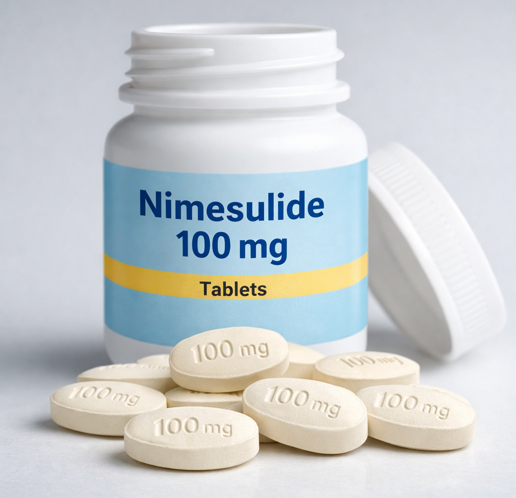
Drugs Safety Alert ResearchNimesulide’s Rise and Reckoning in India: Ending with a Nationwide Ban on... Nimesulide’s Rise and Reckoning in India: Ending with a Nationwide Ban on... Read Post »
Health Tidings ResearchFDA Issues Third CRL for Outlook Therapeutics’ ONS-5010/LYTENAVA in W... FDA Issues Third CRL for Outlook Therapeutics’ ONS-5010/LYTENAVA in W... Read Post »
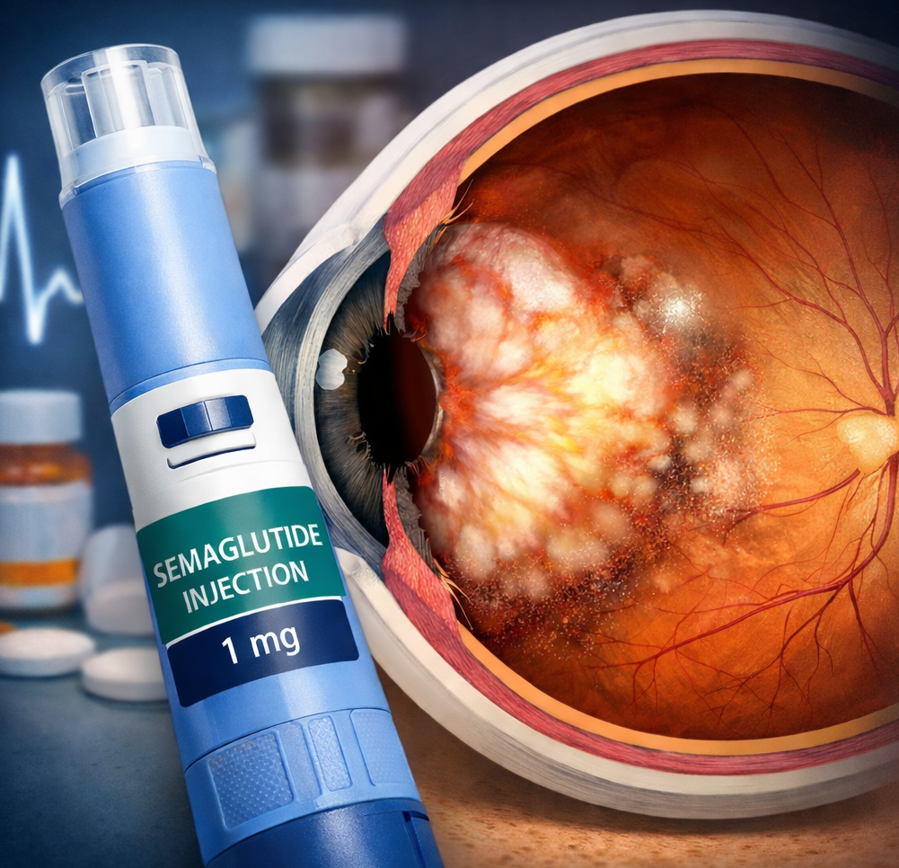
ResearchSemaglutide and Eye Health: A Look at NAION Risk Semaglutide and Eye Health: A Look at NAION Risk Read Post »
Health Tidings ResearchCorcept Therapeutics Receives FDA Complete Response Letter for Relacorilant... Corcept Therapeutics Receives FDA Complete Response Letter for Relacorilant... Read Post »
Health Tidings ResearchGenmab Halts Clinical Development of Acasunlimab After Portfolio Review, Sh... Genmab Halts Clinical Development of Acasunlimab After Portfolio Review, Sh... Read Post »
Drugs Safety Alert ResearchFDA Says PFAS Safety in Cosmetics Remains Uncertain Due to Data Gaps FDA Says PFAS Safety in Cosmetics Remains Uncertain Due to Data Gaps Read Post »
New Drug Approval ResearchGrifols’ FESILTY (Fibrinogen): Evidence, Safety, and Clinical Use Explain... Grifols’ FESILTY (Fibrinogen): Evidence, Safety, and Clinical Use Explain... Read Post »
Drugs Safety Alert ResearchPreliminary Safety Update: Pfizer Notifies Hemophilia Community of Death in... Preliminary Safety Update: Pfizer Notifies Hemophilia Community of Death in... Read Post »
Drugs Safety Alert ResearchJAMA Study Analysing 20 Years of U.S. Data Links GLP-1 Drugs With Persisten... JAMA Study Analysing 20 Years of U.S. Data Links GLP-1 Drugs With Persisten... Read Post »
Drugs Safety Alert ResearchFDA Safety Update: Andexxa Risks Outweigh Benefits After Postmarketing Thro... FDA Safety Update: Andexxa Risks Outweigh Benefits After Postmarketing Thro... Read Post »
Health Tidings ResearchFDA Grants Advanced Manufacturing Technology Designation to Astellas’ Mah... FDA Grants Advanced Manufacturing Technology Designation to Astellas’ Mah... Read Post »
Health Tidings ResearchBlueRock Therapeutics’ Bemdaneprocel Earns Pioneering Regenerative Medica... BlueRock Therapeutics’ Bemdaneprocel Earns Pioneering Regenerative Medica... Read Post »
Lifestyle Research WellnessCan Smoking Make Asthma Inhalers Less Effective? Can Smoking Make Asthma Inhalers Less Effective? Read Post »
Health Tidings ResearchNo Patient Identifiers Needed: FDA Removes a Key Barrier to Using Real-Worl... No Patient Identifiers Needed: FDA Removes a Key Barrier to Using Real-Worl... Read Post »
Food Research WellnessLove Tea but Taking Iron Pills? Read This First Love Tea but Taking Iron Pills? Read This First Read Post »
Drugs Safety Alert ResearchWHO Review Shows Strong Evidence That Vaccines Are Not Linked to Autism WHO Review Shows Strong Evidence That Vaccines Are Not Linked to Autism Read Post »
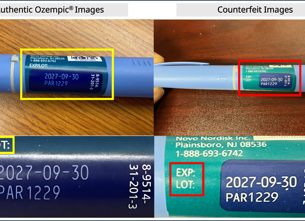
Drugs Safety Alert ResearchFDA Alerts on Fake Ozempic in Legitimate US Supply: Novo Nordisk Guidance A... FDA Alerts on Fake Ozempic in Legitimate US Supply: Novo Nordisk Guidance A... Read Post »
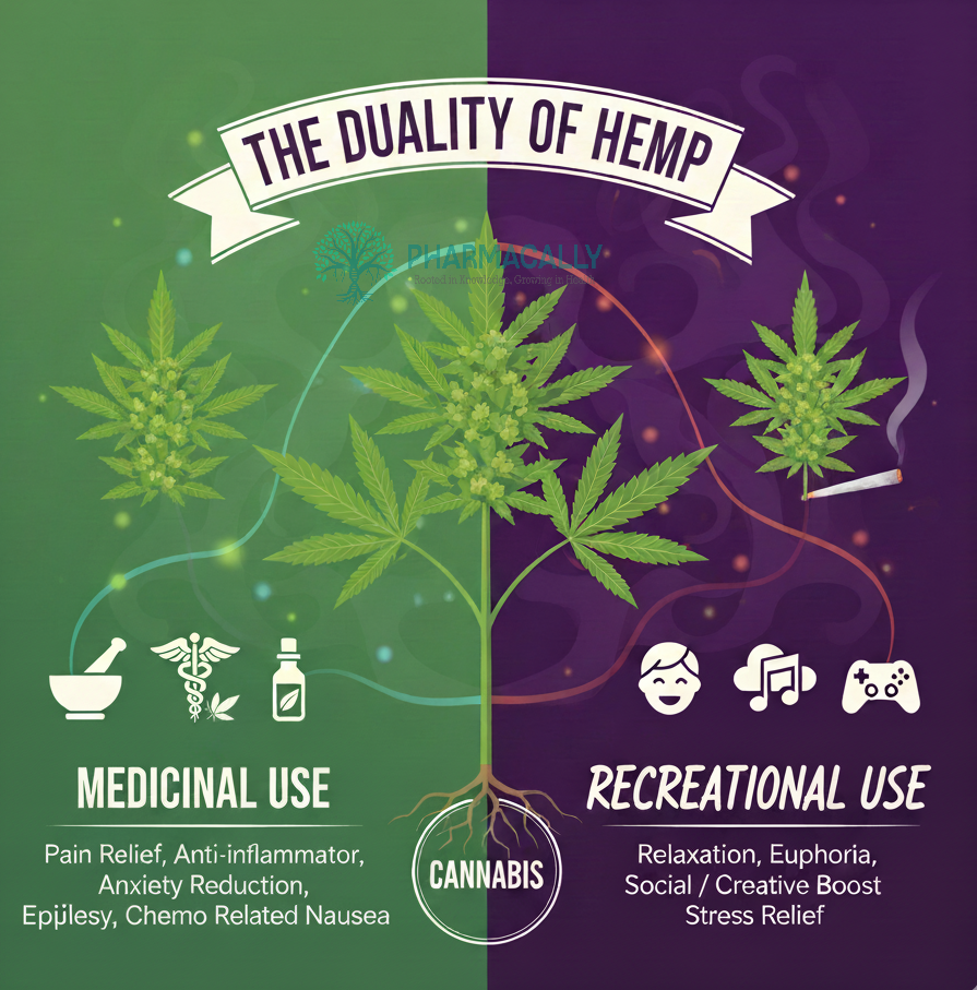
Lifestyle Research WellnessUnderstanding Cannabis: How Medical and Recreational Use Differ in Purpose,... Understanding Cannabis: How Medical and Recreational Use Differ in Purpose,... Read Post »
Health Tidings ResearchFDA Moves Toward Real-World Evaluation of Digital Devices With TEMPO Progra... FDA Moves Toward Real-World Evaluation of Digital Devices With TEMPO Progra... Read Post »
Health Tidings ResearchAbbott Launches Two New Ensure® Max Protein Shakes: 42g Protein and 2-in-1... Abbott Launches Two New Ensure® Max Protein Shakes: 42g Protein and 2-in-1... Read Post »
Drugs Safety Alert ResearchFDA Seizes 73,000 Illegal 7-OH Opioid Products worth $1M in Missouri: Major... FDA Seizes 73,000 Illegal 7-OH Opioid Products worth $1M in Missouri: Major... Read Post »
Drugs Safety Alert ResearchFDA Final Guidance on QTc Information in Human Prescription Drug and Biolog... FDA Final Guidance on QTc Information in Human Prescription Drug and Biolog... Read Post »
Health Tidings ResearchEli Lilly Lowers Single-Dose Zepbound Vial Prices Eli Lilly Lowers Single-Dose Zepbound Vial Prices Read Post »
Drugs Safety Alert ResearchAustralia’s TGA Updates Safety Warnings for GLP-1 Receptor Agonists a... Australia’s TGA Updates Safety Warnings for GLP-1 Receptor Agonists a... Read Post »
ResearchWHO Measles Report 2025: Vaccination Triumphs save Millions but Outbreaks W... WHO Measles Report 2025: Vaccination Triumphs save Millions but Outbreaks W... Read Post »
Drugs Safety Alert ResearchAbbott Initiates Medical Device Correction for Freestyle Libre 3 Sensors in... Abbott Initiates Medical Device Correction for Freestyle Libre 3 Sensors in... Read Post »
Drugs Safety Alert ResearchThe Truth about “Ozempic Penis”: What Men Should Know About Semaglutide... The Truth about “Ozempic Penis”: What Men Should Know About Semaglutide... Read Post »
Health Tidings ResearchEli Lilly Hits $1 Trillion Market Cap: How Mounjaro and Zepbound Revolution... Eli Lilly Hits $1 Trillion Market Cap: How Mounjaro and Zepbound Revolution... Read Post »
Drugs Safety Alert ResearchFDA Investigates Fatal Case of Neutralizing Antibodies to ADAMTS13 after Ad... FDA Investigates Fatal Case of Neutralizing Antibodies to ADAMTS13 after Ad... Read Post »
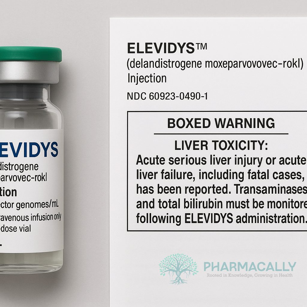
Drugs Safety Alert ResearchSarepta’s ELEVIDYS Updated FDA Labeling: New Boxed Warning and Narrow... Sarepta’s ELEVIDYS Updated FDA Labeling: New Boxed Warning and Narrow... Read Post »
Lifestyle ResearchHow Quitting Tobacco Adds Years to HIV and Tuberculosis Patients’ Lives How Quitting Tobacco Adds Years to HIV and Tuberculosis Patients’ Lives Read Post »
Disease & Drugs Drugs Safety Alert ResearchFDA Revises Menopause Hormone Therapy Labels, Removing Black Box Safety War... FDA Revises Menopause Hormone Therapy Labels, Removing Black Box Safety War... Read Post »
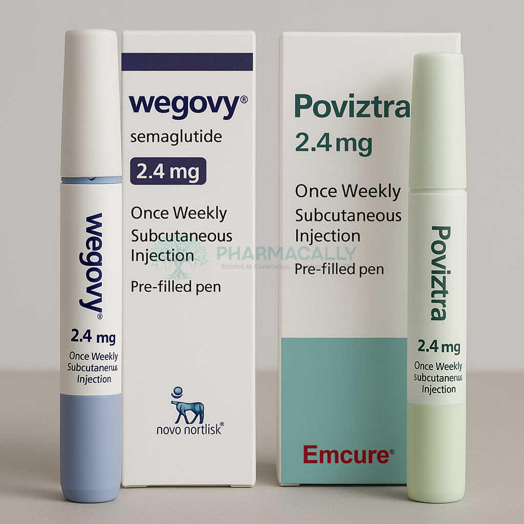
Health Tidings ResearchNovo Nordisk and Emcure Join Forces to Expand Semaglutide Market in India w... Novo Nordisk and Emcure Join Forces to Expand Semaglutide Market in India w... Read Post »
Drugs Safety Alert ResearchFDA Crackdown: 18 Warning Letters Issued Over Illegal Marketing of Unapprov... FDA Crackdown: 18 Warning Letters Issued Over Illegal Marketing of Unapprov... Read Post »
ResearchSemaglutide Users Report Major Reduction in “Food Noise” and Better Men... Semaglutide Users Report Major Reduction in “Food Noise” and Better Men... Read Post »
Drugs Safety Alert ResearchFDA’s New Move to Protect Children from Unapproved Fluoride Drug Prod... FDA’s New Move to Protect Children from Unapproved Fluoride Drug Prod... Read Post »
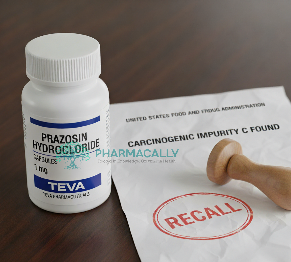
Drugs Safety Alert ResearchTeva Pharmaceuticals Issues Voluntary Recall of Prazosin Hydrochloride Caps... Teva Pharmaceuticals Issues Voluntary Recall of Prazosin Hydrochloride Caps... Read Post »
Drugs Safety Alert ResearchFDA Safety Alert: Increased Reports of Severe Allergic Reactions to Specifi... FDA Safety Alert: Increased Reports of Severe Allergic Reactions to Specifi... Read Post »
Drugs Safety Alert ResearchFDA Tightens Warnings on Tranexamic Acid Amid Deadly Mix-Ups with Spinal An... FDA Tightens Warnings on Tranexamic Acid Amid Deadly Mix-Ups with Spinal An... Read Post »
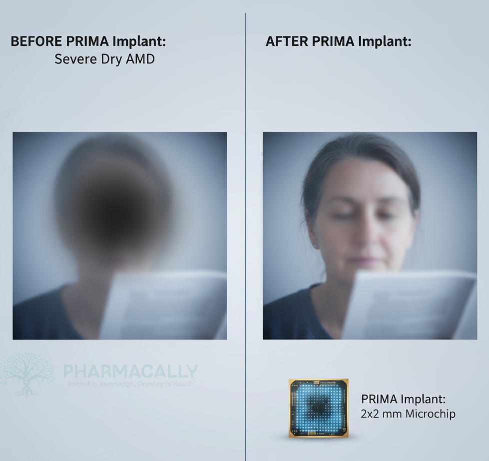
Clinical Trails ResearchFirst Successful Restoration of Central Vision in Dry AMD Using Subretinal ... First Successful Restoration of Central Vision in Dry AMD Using Subretinal ... Read Post »
ResearchWhat the WHO 2025 Antibiotic Resistance Report Warns: The World’s Fight a... What the WHO 2025 Antibiotic Resistance Report Warns: The World’s Fight a... Read Post »