New Drug ApprovalHealth Canada Approves REDEMPLO (plozasiran) for Familial Chylomicronemia S... Health Canada Approves REDEMPLO (plozasiran) for Familial Chylomicronemia S... Read Post »
Health Tidings ResearchAbbott Launches Libre Assist in Libre App to Improve In-the-Moment Food Dec... Abbott Launches Libre Assist in Libre App to Improve In-the-Moment Food Dec... Read Post »
New Drug ApprovalModerna Files With FDA, EMA, Health Canada and TGA for New mRNA Flu Shot Moderna Files With FDA, EMA, Health Canada and TGA for New mRNA Flu Shot Read Post »
Health TidingsWegovy Pill Now Available in the U.S.: First Oral GLP-1 Weight-Loss Medicin... Wegovy Pill Now Available in the U.S.: First Oral GLP-1 Weight-Loss Medicin... Read Post »
New Drug ApprovalAltimmune’s Pemvidutide Granted FDA Breakthrough Therapy Designation for ... Altimmune’s Pemvidutide Granted FDA Breakthrough Therapy Designation for ... Read Post »
New Drug ApprovalGSK’s Nucala (Mepolizumab) Gains China NMPA Approval for Eosinophilic... GSK’s Nucala (Mepolizumab) Gains China NMPA Approval for Eosinophilic... Read Post »
New Drug ApprovalSanofi’s Tzield (Teplizumab) Gains FDA Priority Review for 1-Year-Old... Sanofi’s Tzield (Teplizumab) Gains FDA Priority Review for 1-Year-Old... Read Post »
New Drug ApprovalMHRA Approves Kostaive (Zapomeran) sa-mRNA COVID Booster for UK Adults-Key ... MHRA Approves Kostaive (Zapomeran) sa-mRNA COVID Booster for UK Adults-Key ... Read Post »
New Drug ApprovalUltragenyx Completes Rolling BLA for First-in-Class DTX401 Gene Therapy in ... Ultragenyx Completes Rolling BLA for First-in-Class DTX401 Gene Therapy in ... Read Post »
ResearchPolyrizon Submits Pre-Request for Designation to FDA for PL-16 Viral Blocke... Polyrizon Submits Pre-Request for Designation to FDA for PL-16 Viral Blocke... Read Post »
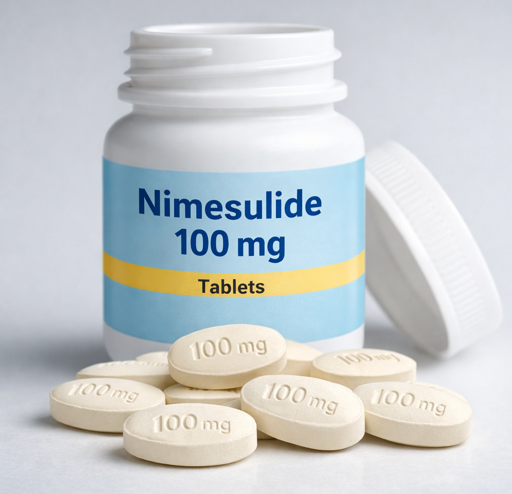
Drugs Safety Alert ResearchNimesulide’s Rise and Reckoning in India: Ending with a Nationwide Ban on... Nimesulide’s Rise and Reckoning in India: Ending with a Nationwide Ban on... Read Post »
Drugs Safety AlertFDA Adds New Safety Warning to ALTUVIIIO Label FDA Adds New Safety Warning to ALTUVIIIO Label Read Post »
New Drug ApprovalFDA Accepts Axsome’s AXS-05 Supplemental NDA and Grants Priority Review f... FDA Accepts Axsome’s AXS-05 Supplemental NDA and Grants Priority Review f... Read Post »
Health Tidings ResearchFDA Issues Third CRL for Outlook Therapeutics’ ONS-5010/LYTENAVA in W... FDA Issues Third CRL for Outlook Therapeutics’ ONS-5010/LYTENAVA in W... Read Post »
ResearchSemaglutide and Eye Health: A Look at NAION Risk Semaglutide and Eye Health: A Look at NAION Risk Read Post »
Health Tidings ResearchCorcept Therapeutics Receives FDA Complete Response Letter for Relacorilant... Corcept Therapeutics Receives FDA Complete Response Letter for Relacorilant... Read Post »
Health TidingsNovo Nordisk Wins Final Patent Ruling on Semaglutide in China Novo Nordisk Wins Final Patent Ruling on Semaglutide in China Read Post »
New Drug ApprovalFDA Clears NEREUS, First New Motion Sickness Drug in Decades FDA Clears NEREUS, First New Motion Sickness Drug in Decades Read Post »
Clinical TrailsDenali’s Tividenofusp Alfa (DNL310) Phase 1/2 Data in Hunter Syndrome wit... Denali’s Tividenofusp Alfa (DNL310) Phase 1/2 Data in Hunter Syndrome wit... Read Post »
New Drug ApprovalUnicycive Resubmits NDA for Oxylanthanum Carbonate for Hyperphosphatemia Ma... Unicycive Resubmits NDA for Oxylanthanum Carbonate for Hyperphosphatemia Ma... Read Post »
New Drug ApprovalFDA Grants Breakthrough Therapy to Ulixacaltamide (PRAX-944) for Essential ... FDA Grants Breakthrough Therapy to Ulixacaltamide (PRAX-944) for Essential ... Read Post »
Policy & AcquisitionsJohnson & Johnson Finalizes Acquisition of Halda Therapeutics Johnson & Johnson Finalizes Acquisition of Halda Therapeutics Read Post »
Health Tidings ResearchGenmab Halts Clinical Development of Acasunlimab After Portfolio Review, Sh... Genmab Halts Clinical Development of Acasunlimab After Portfolio Review, Sh... Read Post »
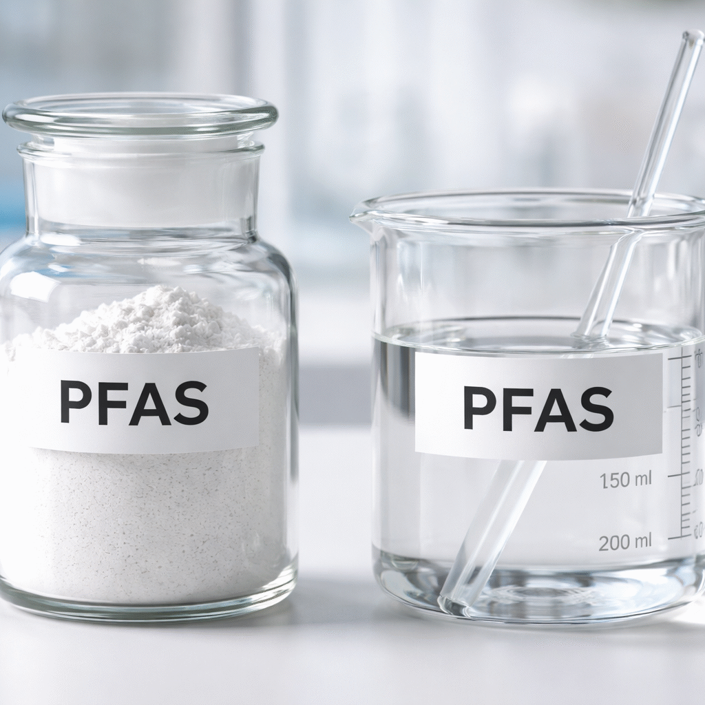
Drugs Safety Alert ResearchFDA Says PFAS Safety in Cosmetics Remains Uncertain Due to Data Gaps FDA Says PFAS Safety in Cosmetics Remains Uncertain Due to Data Gaps Read Post »
New Drug ApprovalFirst-in-Class TRPV1 Inhibitor Avarept Approved in Japan for Dry Eye Diseas... First-in-Class TRPV1 Inhibitor Avarept Approved in Japan for Dry Eye Diseas... Read Post »
Food WellnessDoes Yogurt Prevent Antibiotic-Associated Diarrhea? What the Research Shows Does Yogurt Prevent Antibiotic-Associated Diarrhea? What the Research Shows Read Post »
New Drug Approval ResearchGrifols’ FESILTY (Fibrinogen): Evidence, Safety, and Clinical Use Explain... Grifols’ FESILTY (Fibrinogen): Evidence, Safety, and Clinical Use Explain... Read Post »
Clinical TrailsGalapagos GLPG3667 Meets Primary Endpoint in Dermatomyositis Phase 3-Enabli... Galapagos GLPG3667 Meets Primary Endpoint in Dermatomyositis Phase 3-Enabli... Read Post »
Clinical TrailsTirzepatide Matches Dulaglutide on Major Cardiovascular Outcomes in SURPASS... Tirzepatide Matches Dulaglutide on Major Cardiovascular Outcomes in SURPASS... Read Post »
Health TidingsEnvafolimab Granted FDA Orphan Drug Designation for Gastric and GEJ Cancer:... Envafolimab Granted FDA Orphan Drug Designation for Gastric and GEJ Cancer:... Read Post »
Clinical TrailsJohnson and Johnson’s JNJ-5939 Falls Short in DUPLEX-AD Phase 2b Trial fo... Johnson and Johnson’s JNJ-5939 Falls Short in DUPLEX-AD Phase 2b Trial fo... Read Post »
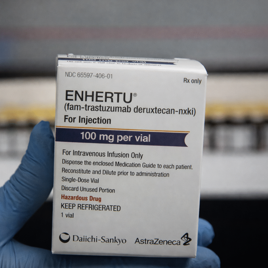
New Drug ApprovalENHERTU Approved in China for HER2-Low and HER2-Ultralow Metastatic Breast ... ENHERTU Approved in China for HER2-Low and HER2-Ultralow Metastatic Breast ... Read Post »
New Drug ApprovalFDA Approves YARTEMLEA: First TA-TMA Therapy Post-Stem Cell Transplant FDA Approves YARTEMLEA: First TA-TMA Therapy Post-Stem Cell Transplant Read Post »
Clinical TrailsBiogen Reports Final VALOR Results Showing Earlier QALSODY Treatment May Sl... Biogen Reports Final VALOR Results Showing Earlier QALSODY Treatment May Sl... Read Post »
New Drug ApprovalIncyte’s Zynyz Receives First Approval in Japan for Advanced SCAC Incyte’s Zynyz Receives First Approval in Japan for Advanced SCAC Read Post »
New Drug ApprovalJapan Expands Pediatric Asthma Treatment with Dupixent Approval Japan Expands Pediatric Asthma Treatment with Dupixent Approval Read Post »
Clinical TrailsNeurocrine’s Phase 3 KINECT-DCP Trial of Valbenazine Fails to Meet En... Neurocrine’s Phase 3 KINECT-DCP Trial of Valbenazine Fails to Meet En... Read Post »
Policy & AcquisitionsSanofi to Acquire Dynavax, Strengthening Adult Vaccine Portfolio with HEPLI... Sanofi to Acquire Dynavax, Strengthening Adult Vaccine Portfolio with HEPLI... Read Post »
Health TidingsFDA Issues CRL for Sanofi’s Tolebrutinib in Non-Relapsing Secondary Progr... FDA Issues CRL for Sanofi’s Tolebrutinib in Non-Relapsing Secondary Progr... Read Post »
New Drug ApprovalZai Lab Wins China Approval for COBENFY, a First-in-Class Muscarinic Therap... Zai Lab Wins China Approval for COBENFY, a First-in-Class Muscarinic Therap... Read Post »
New Drug ApprovalU.S. FDA Approves Agios’ AQVESME (Mitapivat) as First Treatment for Anemi... U.S. FDA Approves Agios’ AQVESME (Mitapivat) as First Treatment for Anemi... Read Post »
Wellness Women & Child HealthHydration and UTIs: How Drinking Water Supports Antibiotic Treatment Hydration and UTIs: How Drinking Water Supports Antibiotic Treatment Read Post »
Drugs Safety Alert ResearchPreliminary Safety Update: Pfizer Notifies Hemophilia Community of Death in... Preliminary Safety Update: Pfizer Notifies Hemophilia Community of Death in... Read Post »
New Drug ApprovalJapan Approves Finerenone for Chronic Heart Failure with Preserved or Mildl... Japan Approves Finerenone for Chronic Heart Failure with Preserved or Mildl... Read Post »
New Drug ApprovalEuropean Commission Approves TREMFYA® (guselkumab) for Paediatric Plaque P... European Commission Approves TREMFYA® (guselkumab) for Paediatric Plaque P... Read Post »
New Drug ApprovalNovo Nordisk’s Wegovy Pill Wins FDA Approval Following OASIS Trial Result... Novo Nordisk’s Wegovy Pill Wins FDA Approval Following OASIS Trial Result... Read Post »
New Drug ApprovalSubcutaneous Lunsumio VELO™ Approved: 75% ORR in Heavily Pretreated Folli... Subcutaneous Lunsumio VELO™ Approved: 75% ORR in Heavily Pretreated Folli... Read Post »
Drugs Safety Alert ResearchJAMA Study Analysing 20 Years of U.S. Data Links GLP-1 Drugs With Persisten... JAMA Study Analysing 20 Years of U.S. Data Links GLP-1 Drugs With Persisten... Read Post »
Health TidingsIpsen licenses SIM0613 ADC from Simcere Zaiming, targeting Phase I in 2026 Ipsen licenses SIM0613 ADC from Simcere Zaiming, targeting Phase I in 2026 Read Post »
Health TidingsJ&J Supports HRS Registry to Advance PFA Evidence in AFib J&J Supports HRS Registry to Advance PFA Evidence in AFib Read Post »