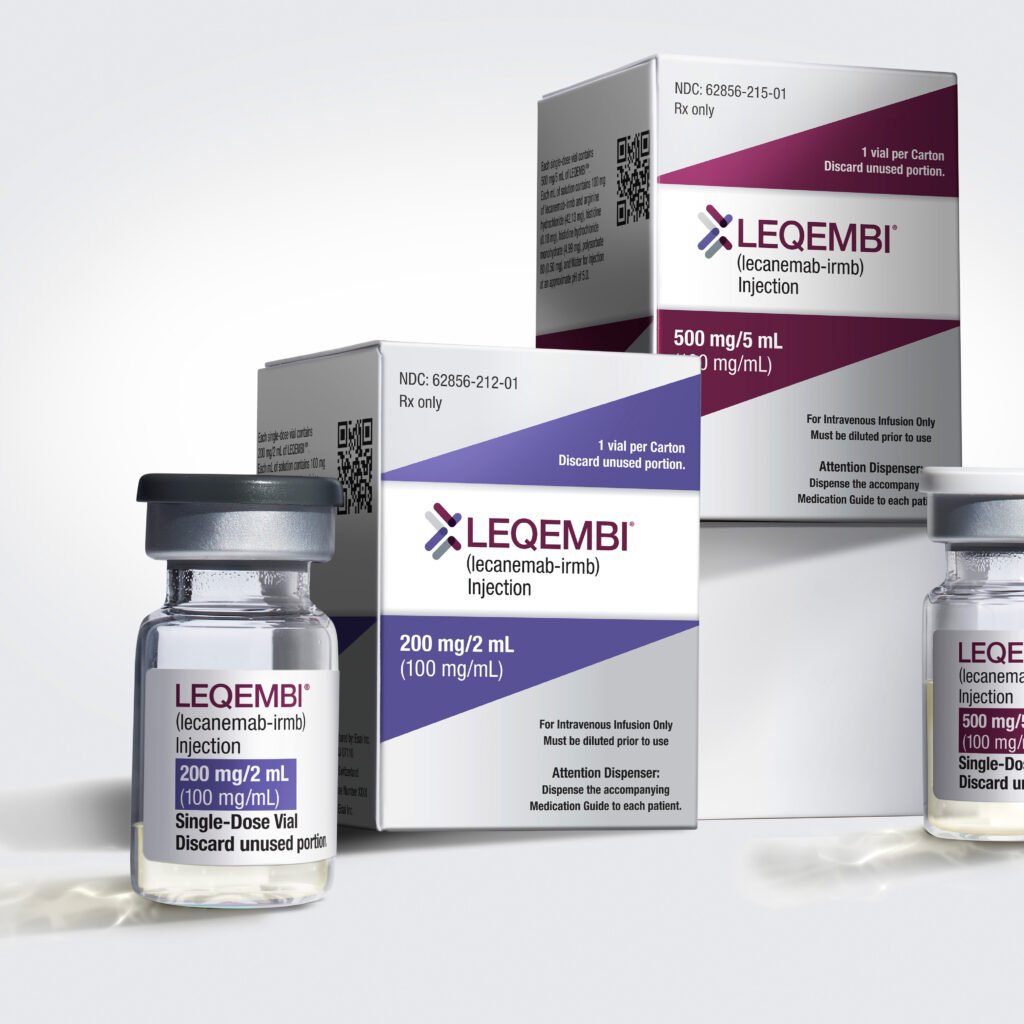
pexels-charliehelenrobinson-4531306

Wrong Blood, Lost Life: How a Blood Transfusion Error at SMS Hospital, Rajasthan Sparked a State-wide Medical Reckoning

Written and Reviewed by Vikas Londhe M.Pharm (pharmacology)

Assisted By: Shakuntala Kawhale (M.Pharm pharmacology)

pexels-charliehelenrobinson-4531306
Source: Pexels.com

On May 20, 2025, a 23-year-old pregnant woman died in Jaipur’s prestigious Sawai Man Singh (SMS) Hospital, not from the illness she was fighting but from a blood transfusion mistake that was preventable and never should have happened.

A female patient from Tonk district, who was seven months pregnant, was admitted to SMS Hospital on May 12 with severe anaemia and miliary tuberculosis. Her pregnancy was already classified as high-risk. However, she was mistakenly given A+ blood, despite her actual blood type being B+.

The error was not caught until it was too late. By the time symptoms like fever, chills, hematuria, and rapid heart rate appeared, around 350 ml of incompatible blood had already been transfused. Patient succumbed to the reaction later that night.

Timeline of Errors

The tragedy was not the result of a single misstep but a chain of preventable lapses:

  •  No dual verification between the blood samples sent from the ward and the patient’s records.
  • Incorrect blood group identification during ward-level preparation.
  • Failure to match identifiers (yellow tag, ABHA ID, blood bag barcode) at the point of transfusion.
  • Inadequate monitoring in the critical first 15–30 minutes post-transfusion, when early signs of reaction generally appear.
  • No immediate escalation occurred when the patient developed symptoms, delaying corrective measures.
  • Delayed and incomplete communication with her family, who were not immediately informed of the transfusion error.

This was the third transfusion-related death involving SMS and its affiliated hospitals in just over a year, highlighting a pattern of systemic failure.

What the Internal Inquiry Found and Why It Was Rejected

SMS Hospital conducted an internal investigation and acknowledged the transfusion error. However, the report controversially claimed that the mismatched blood was not the direct cause of death, pointing towards the patient’s tuberculosis and low haemoglobin levels.

This standpoint received immediate criticism from many levels. The Rajasthan government rejected the internal findings, labelled them unsatisfactory and ordered an independent, high-level investigation. Health Minister Gajendra Singh Khimsar announced the formation of a committee comprising senior doctors, administrative officials, and public health experts, tasked with submitting a report within three days.

The case also reached the Rajasthan State Human Rights Commission, which issued a notice demanding detailed reports by June 12.

Rajasthan Government’s Official Response: a Shift toward Safety

Under the public pressure and clear protocol violations, the Rajasthan government took immediate and serious action:

Key Policy Decisions Announced

Digital Blood Group mapping

All patients in state hospitals will now have their blood type linked to their Ayushman Bharat Health Account ID (ABHA ID), reducing room for clerical or manual entry errors.

Compulsory Admission—Time Blood Typing

Every patient must now undergo blood grouping at admission, with results entered into a centralized digital lab system.

Dual Verification at Blood Banks

Before releasing any blood unit, technicians must re-verify the patient’s blood type and documentation, independent of the ward-level grouping.

Detailed Chain of Custody Documentation

Blood request forms must now contain:

Name and mobile number of the requesting resident

Attending the duty doctor’s name

Name of the staff that collected the sample and the blood bag

Standardized Monitoring Protocol during Transfusion

Clinical staff must now:

Match the patient ID, yellow wristband, and blood label

Monitor vitals at 0, 5, 15, and 30 minutes during the transfusion

Record all observations in real time

Statewide SOP Enforcement across RMES Hospitals

SMS-level safety protocols are now mandatory across all government medical colleges and district hospitals under the Rajasthan Medical Education Society (RMES).

Patient Safety Lessons: When Systems Fail, People Pay the Price

The loss of life is a staggering reminder of how even routine clinical procedures can turn deadly without robust precaution. Blood transfusions are considered high-alert medical procedures, requiring multiple fail-safe mechanisms to prevent mismatches. When these mechanisms are skipped whether due to staff shortages, lack of training, or system fatigue the consequences are often fatal.

This case illustrates the urgent need for error-proof processes, accountability tracking, and a culture where speaking up about unsafe practices is encouraged.

Pharmacally’s Take: Reform Begins with Acknowledging the Risk

While the Rajasthan government’s response has been proactive and progressive, the real test lies in enforcement. Policies on paper must translate to practice on the ground. Frontline workers residents, nurses, lab techs must be trained, audited, and supported in following these updated protocols.

At Pharmacally.com, we advocate for healthcare systems that:

  • Prioritize patient safety
  • Embed digital tools to reduce human error.
  • Enforce chain-of-responsibility in critical procedures, and
  • Promote transparent investigations when outcomes go wrong.

Final Word: From Tragedy to Transformation

This patient should have gone home with her newborn in a few months. Instead, her death has exposed deep vulnerabilities in our public health system. If Rajasthan succeeds in turning this tragedy into a safety revolution, it could become a blueprint for the rest of India.

References:

Gandhi a, görlinger k, nair sc, kapoor pm, trikha a, mehta y, handoo a, karlekar a, kotwal j, john j, apte s. Patient blood management in india-review of current practices and feasibility of applying appropriate standard of care guidelines. A position paper by an interdisciplinary expert group. Journal of anaesthesiology clinical pharmacology. 2021 jan 1;37(1):3-13.

Bisht a, singh s, marwaha n. Hemovigilance program-india. Asian journal of transfusion science. 2013 jan 1;7(1):73-4.

Mammen jj, asirvatham es, sarman cj, ranjan v, charles b. A review of legal, regulatory, and policy aspects of blood transfusion services in india: issues, challenges, and opportunities. Asian journal of transfusion science. 2021 jul 1;15(2):204-11

Rajasthan to streamline blood transfusion system in all hospitals, News Arena India, https://newsarenaindia.com/nation/rajasthan-to-tighten-hospital-blood-transfusion-rules/45595

State Gov to tightens blood safety protocols after transfusion deaths, times of India, https://timesofindia.indiatimes.com/city/jaipur/state-govt-to-tighten-blood-safety-protocols-after-transfusion-death/articleshow/121421643.cms

Rajasthan hospital accused of fatal transfusion error after pregnant woman, 23, dies, The Economic Times, https://economictimes.indiatimes.com/news/india/rajasthan-hospital-accused-of-fatal-transfusion-error-after-pregnant-woman-23-dies/articleshow/121362945.cms?from=mdr

2149005991

Mandukaparni (Centella asiatica): The Ayurvedic Herb That Rewires Your Brain & Rejuvenates Your Body

Written By: Lavanya Chavhan, B.Pharm

Reviewed By: Ayurvedacharya Dr. Gaurav Pathare, BAMS

2149005991

Mandukaparni (Gotu Kola) is an ancient Ayurvedic herb known for boosting memory, reducing anxiety, and healing the skin. Backed by clinical evidence and meta-analysis, learn how to use Mandukaparni effectively for optimal health

At Pharmacally, we believe that the best medicine combines ancient wisdom with modern science. One such powerful herb is Mandukaparni a small green leaf with big healing potential.

If you’re dealing with brain fog, skin problems, stress, or slow-healing wounds, Mandukaparni might be the natural support your body needs.

Known scientifically as Centella asiatica (CA), also known as Mandukaparni, Indian pennywort, or Gotu Kola, Mandukaparni has been a part of Ayurvedic medicine for centuries. In ancient texts, it’s described as a “Medhya Rasayana” an herb that nourishes and sharpens the mind. In Sanskrit Manduk means frog and Parni means leaves; which means shape of the leaves are resembled with the shape of feets of the frog, hence Mandukaparni.

Traditionally used in India and Southeast Asia, this herb is now being explored in modern clinical trials and meta-analyses, confirming many of its traditional uses. From boosting brain function to improving skin repair, Mandukaparni is gaining recognition as a truly holistic healer. It is mentioned in ancient texts like the Sushruta Samhita. The herb is also traditionally used in Java, Indonesia, and China, where it is considered a “miracle elixir of life.”

Active constituents involved in pharmacological action of a Mandukaparni

At the heart of its therapeutic power lies a complex array of phytochemicals, natural compounds that work in synergy to promote healing.

At the core of Mandukaparni’s healing power lies a unique group of compounds known as triterpenoid saponinsThe most studied are asiaticoside and madecassoside. These compounds are primarily responsible for promoting wound healing by stimulating collagen synthesis and reducing inflammation.

In addition to triterpenoids, Mandukaparni contains significant amounts of flavonoids and polyphenols such as quercetin, kaempferol, and rutin. These compounds contribute to the herb’s potent antioxidant activity, helping to neutralize free radicals and protect brain cells from oxidative stress. Studies suggest that these flavonoids work in synergy with triterpenoids to enhance cognitive functions and delay age-related neurological decline.

Mandukaparni also yields a variety of essential oils, obtained through steam distillation of its leaves. Key volatile compounds include caryophyllene, pinene, and humulene, each of which exhibits antimicrobial, anti-inflammatory, and mood-stabilizing properties. These oils are often used in skin creams, ointments, and aromatherapy preparations for added therapeutic benefit, particularly in wound care. Further phytochemical screening reveals the presence of β-sitosterol, a plant sterol known for supporting cardiovascular health and cholesterol balance.

Health Benefits of Mandukaparni

Sharpens Memory & Boosts Mental Clarity

Mandukaparni is widely recognized as a natural brain tonic. It is believed to nourish the nervous system, support cognitive functions, and promote mental clarity. Regular use is said to enhance memory retention, sharpen focus, and reduce mental fatigue, making it a popular choice among students and individuals under high mental stress.

Reduces Stress & Calms the Mind

This herb is often used to soothe anxiety, restlessness, and irritability. In Ayurvedic texts, Mandukaparni is said to balance the “vata” dosha, which governs the nervous system. It promotes a calm, grounded mental state and helps reduce symptoms of stress-induced imbalances such as insomnia, overthinking, and emotional instability.

Revitalizes Skin Health

Mandukaparni has long been used to improve skin texture, tone, and elasticity. It is considered excellent for managing various skin disorders including dryness, discoloration, and minor wounds. Its regenerative properties support faster healing of cuts, scars, and burns while rejuvenating the skin from within.

Supports Cardiovascular and Circulatory Health

Centella asiatica is believed to strengthen blood vessels and promote better circulation. It may help relieve symptoms of varicose veins, swelling in the legs, and heaviness caused by poor venous return. By improving blood flow, it nourishes tissues and supports overall vascular health.

Supports Healthy Digestion

Mandukaparni is traditionally used to soothe and strengthen the digestive tract. It may reduce inflammation in the gut lining and improve digestion by calming excess heat and acidity. It’s also believed to promote better assimilation of nutrients and relieve minor digestive discomfort.

Promotes Longevity & Rejuvenation

In Ayurveda, Mandukaparni is regarded as a rasayana a substance that promotes longevity, vitality, and youthfulness. It is believed to rejuvenate tissues, slow down the aging process, and maintain balance in the body’s natural systems. Regular use is said to support energy levels, immunity, and overall well-being.

Improves Hair and Scalp Health

Applied externally or taken internally, Mandukaparni is beneficial for hair strength and scalp nourishment. It is traditionally used to reduce hair fall, promote hair growth, and combat dandruff. Its cooling nature helps soothe inflamed or itchy scalps, promoting a healthy hair environment.

Supports Joint Flexibility & Tissue Repair

The herb is believed to support flexibility and joint mobility. It helps nourish connective tissues, strengthen cartilage, and reduce stiffness, making it useful for people experiencing age-related wear and tear or joint discomfort.

How to Take Mandukaparni for Best Results

Form

Dosage

Usage

Capsules/Tablets

300–600 mg per day

With water after meals

Powder (Churna)

1–2 grams daily

Mix with warm water, honey, or ghee

Decoction (Kashayam)

30–50 ml

Once or twice daily

Tea

1–2 cups/day

Boil leaves for 10 minutes

Topical Creams

As directed

For wounds, scars, and skin rejuvenation

Note: Always consult your healthcare practitioner or Ayurvedic expert before starting any new herb, especially if you are pregnant, nursing, or taking medications.

Conclusion

Mandukaparni (Centella asiatica) stands as a timeless herbal treasure that gently nurtures the body, mind, and spirit. From sharpening memory to calming the nervous system, revitalizing the skin, supporting circulation, and rejuvenating the joints, its wide-reaching benefits are a testament to its revered status in Ayurveda as a medhya rasayana and rasayana a promoter of both intellect and longevity.

What gives Mandukaparni its unique healing potential is not just its traditional value, but also its rich phytochemical profile. It contains powerful bioactive compounds such as asiaticoside, madecassoside, asiatic acid, and madecassic acid. These triterpenoids are known to support tissue regeneration, enhance collagen synthesis, reduce inflammation, and promote neuroprotection at a cellular level. Additionally, the presence of flavonoids and saponins further enhances its antioxidant and adaptogenic properties.

In essence, Mandukaparni is more than a medicinal herb it is a holistic ally for modern wellness. Whether you’re seeking clarity of mind, youthful skin, restful sleep, or internal balance, this humble green leaf has something to offer.

References

Boju Sun, Lily Wu, You Wu et al, Therapeutic Potential of Centella asiatica and Its Triterpenes: A Review, Front. Pharmacol, 04 September 2020 Sec. Ethnopharmacology Volume 11 – 2020 | https://doi.org/10.3389/fphar.2020.568032

Oluranti Mopelola Lawal, Fatima Wakel, Matthijs Dekker, Consumption of fresh Centella asiatica improves short term alertness and contentedness in healthy females, Journal of Functional Foods, Volume 77, 2021, 104337, https://doi.org/10.1016/j.jff.2020.104337.

Lokanathan Y, Omar N, Ahmad Puzi NN, Saim A, Hj Idrus R. Recent Updates in Neuroprotective and Neuroregenerative Potential of Centella asiatica. Malays J Med Sci. 2016 Jan;23(1):4-14. PMID: 27540320; PMCID: PMC4975583.

Eduviere, A. T., Awhin, P. E., Edje, K. E., Otomewo, L. O., Adeoluwa, O. A., & Winter, J. E. (2021). Adaptogenic potential of Centella lujica supplement in sleep deprived mice. International Journal of Research in Medical Sciences9(11), 3269–3276. https://doi.org/10.18203/2320-6012.ijrms20214408

Jeevan Chandra, Himanshu Joshi, Pankaj Bahuguna, Anti-stress effect of Centella asiatica in rats, Sch. Acad. J. Biosci., 2015; 3(8):668-675, DOI : 10.36347/sajb.2015.v03i08.005

Gohil KJ, Patel JA, Gajjar AK. Pharmacological Review on Centella asiatica: A Potential Herbal Cure-all. Indian J Pharm Sci. 2010 Sep;72(5):546-56. doi: 10.4103/0250-474X.78519. PMID: 21694984; PMCID: PMC3116297.

Health Benefits of Mandukaparni (Centella Asiatica), https://www.all-cures.com/cure/5451-Health-Benefits-of-Mandukaparni-(Centella-Asiatica)

Tan, S.C.; Bhattamisra, S.K.; Chellappan, D.K.; Candasamy, M. Actions and Therapeutic Potential of Madecassoside and Other Major Constituents of Centella asiatica: A Review. Appl. Sci. 2021, 11, 8475. https://doi.org/10.3390/app11188475

Razali NNM, Ng CT, Fong LY. Cardiovascular Protective Effects of Centella asiatica and Its Triterpenes: A Review. Planta Med. 2019 Nov;85(16):1203-1215. doi: 10.1055/a-1008-6138. Epub 2019 Sep 20. PMID: 31539918.

Dora Bhavna, Khatri Jyoti, Centella Asiatica: the elixir of life, International Journal of Research in Ayurveda & Pharmacy, 2(2), 2011 431-438

19648

“Deadly Saline Killed 8: Neuromelioidosis Outbreak in Tamil Nadu Linked to Contaminated Dental Saline – What Went Wrong?”

Written by: Dr. Arshada Fathin, PharmD (Coimbatore, Tamil Nadu)

Reviewed By: Dr. Seema Satbhai (BAMS, MPH, PhD, Public Health)

19648

Neuromelioidosis, a central nervous system manifestation of melioidosis caused by Burkholderia pseudomallei, has emerged as a serious health concern in Vaniyambadi, Tirupattur district, Tamil Nadu, India. An unusual cluster of cases was identified between July 2022 and April 2023. Importantly, 10 out of 21 patients had undergone some dental procedure, most at the same dental clinic. Out of these 10 cases, 8 patients succumbed to death. This has raised critical questions regarding infection control practices in dental care settings. This article provides an overview of the outbreak, disease pathology, diagnostic considerations, and the role of procedural lapses in precipitating iatrogenic transmission.

Background of the Case

The outbreak of neuromelioidosis in Tamil Nadu centres on a cluster of 21 cases reported between July 2022 and April 2023. These included 11 sporadic infections and 10 distinct sub-cluster epidemiologically linked to a dental clinic in Vaniyambadi, Tirupattur district. The affected patients who visited the clinic presented with severe symptoms shortly after dental treatment.

About Neuromelioidosis

Neuromelioidosis results from infection of the central nervous system by Burkholderia pseudomallei, manifesting primarily as brainstem syndrome. Typical clinical features include facial pain, cranial nerve dysfunction, abscess formation, and necrotizing encephalitis. The infection often spreads along neuronal pathways, such as the trigeminal nerve, leading to rapid neurological decline. Adults within the age range of 26 to 44 years are predominantly affected in endemic regions. Diagnosis relies on isolation of the organism from blood, cerebrospinal fluid (CSF), or affected tissues through culture or polymerase chain reaction (PCR) assays. Molecular tools, including WGS and detection of virulence-associated genes such as bimA, aid in strain typing and epidemiological investigations. Management requires prompt initiation of intravenous antibiotics, commonly ceftazidime or meropenem, followed by prolonged oral eradication therapy, i.e., Co-Trimoxazole, for several months. Delay in recognition and treatment often results in poor clinical outcomes, sometimes death.

The Culprit: Burkholderia pseudomallei

This bacterium typically inhabits soil and water, but in this outbreak, it entered patients directly along nerve pathways by contact with mucous membranes in the mouth during dental treatments. This neurological route led to rapid brain infections rather than the more typical bloodstream spread.

Case Description and Microbiological Findings

The outbreak involved 21 patients with neuromelioidosis in Tamil Nadu, with a median age of 33 years, affecting both males and females. Clinical presentations include facial cellulitis, lymphadenopathy, brainstem involvement, and rapid neurological decline. Among the 10 patients with documented dental procedure exposure, 8 succumbed to the illness within a median period of 17 days post-exposure, indicating a higher fatality rate compared to sporadic cases. The high fatality of the outbreak is indicative of an iatrogenic source of infection. A high-level, authoritative investigation was warranted on an immediate basis.

Investigation by Researchers

It all started when a married couple presented at CMC Vellore with acute-onset fever followed by symptoms of brainstem syndrome. Radiological imaging revealed necrotizing brainstem encephalitis, a finding consistent with neuromelioidosis. The relative reported that both patients had undergone a dental procedure at the dental clinic in Vaniyambadi shortly before illness.

A highly authoritative investigation was carried out which led by researchers from CMC Vellore, ICMR-National Institute of Epidemiology, Chennai, and the Tamil Nadu Directorate of Public Health and Preventive Medicine. The primary objective of the investigation was to understand how the infection spread and confirm whether the source was iatrogenic. The outbreak investigation utilized clinical evaluation, microbiological cultures, environmental sampling and whole-genome sequencing (WGS) of bacterial strain to establish the transmission dynamics and identify the source of infection. The initial investigation pointed to the cases from Tirupattur district experiencing an outbreak after undergoing invasive dental procedures at a local dental clinic and suggested possible contamination during dental procedures.

Identified Deviations in Infection Control – Medication Error and Sterility Breach

Based on an interview with the dentist and investigation findings, the dental clinic involved in the outbreak provided a wide range of services, including fixation of partial dentures, full-mouth prophylaxis, root canal treatments, and tooth extraction. The clinic was staffed by a dentist, nursing personnel, and a receptionist; however, none of the staff had formal training in hospital infection control practices.

Normal saline was supplied in sterile 500 mL plastic bottles and used both for wound irrigation during surgical procedures and for dilution of local anesthetic for infiltration. It was observed that the sterile saline bottles were opened using a non-sterile periosteal elevator. These bottles were then loosely resealed and reused over several days until empty. There was no proper sterilization of equipment between patients.

Such practices deviated significantly from established infection control standards. The repeated use of opened saline bottles without proper aseptic technique, combined with the use of non-sterile instruments to open them, increased the risk of contamination. Indeed, an increase in failed dental procedures and complications was reported concurrent with the emergence of neuromelioidosis cases in the district.

Microbiological analysis supported these findings, as Burkholderia pseudomallei was isolated from one of the in-use saline bottles collected during environmental sampling at the clinic. These breaches in sterile technique and medication handling directly contributed to the iatrogenic transmission of the pathogen to patients during dental procedures.

Recommended Preventive Measures

Based on the outbreak findings, several corrective measures are essential to prevent similar incidents in the future:

Strict use of sterile supplies: Single-use sterile saline bottles should be opened aseptically and discarded after one use. The reuse of opened bottles must be avoided.

Adherence to aseptic technique: All dental procedures must be performed under sterile conditions, employing sterilized instruments and protective barriers such as sterile gloves.

Comprehensive staff training: Dental clinic personnel should receive ongoing training in infection prevention and control protocols, emphasizing proper handling of sterile supplies.

Environmental monitoring: Routine microbial testing of dental unit water lines and fluids should be conducted, particularly in regions endemic for B. pseudomallei.

Immediate investigation of infection clusters: Any unusual increase in post-procedural infections must prompt epidemiological investigation and suspension of implicated materials or practices.

Implementing these measures will reduce the risk of iatrogenic infection and ensure patient safety during dental procedures.

Conclusion: A Wake-Up Call for Infection Control in Outpatient Care

The tragic Neuromelioidosis outbreak in Tamil Nadu is more than just a public health incident. It is a warning about how single lapse in clinical hygiene, such as reusing a contaminated saline bottle, can escalate into a fatal outbreak of a rare and deadly infection. While melioidosis is typically associated with environmental exposure in endemic regions, this event underscores an alarming reality: modern medical settings are not immune to such threats when basic infection control protocols are neglected.

The Vaniyambadi dental clinic case highlights critical vulnerabilities in outpatient care, particularly in resource-limited settings where practices like reusing consumables or improper instrument handling may still occur. In this instance, the use of an unsterile periosteal elevator to repeatedly open a saline bottle facilitated the direct introduction of Burkholderia pseudomallei, a soil-dwelling microorganism, into the oral mucosa, bypassing traditional routes of transmission. The result was an outbreak of neuromelioidosis, a rare neurological manifestation with devastating consequences, including an 80% fatality rate among exposed patients.

The rapid identification of the outbreak source, coordinated by CMC Vellore, ICMR-NIE Chennai, and state health authorities, demonstrates the importance of multisectoral collaboration in outbreak investigation.

For healthcare systems across India and globally, this event serves as a wake-up call. It emphasizes the urgent need to:

Reinforce standard infection prevention protocols in dental and outpatient clinics

Mandate the single-use of consumables like saline bottles

Educate practitioners on rare but deadly infections like melioidosis

Improve clinical waste management and sterilization practices

Establish local microbiology networks for early pathogen detection

Ultimately, this outbreak is an earnest reminder that patient safety must never be compromised for convenience. The lives lost in Vaniyambadi are a tragic testament to what can go wrong, but also a call to action, urging the healthcare community to uphold the highest standards of hygiene, vigilance, and accountability.

This investigation and findings are published in the Lancet Regional Health – Southeast Asia

References

Angel Miraclin Thirugnanakumar, Prabu Rajkumar, Karthik Gunasekaran et al, Neuromelioidosis outbreak in Tamil Nadu, India: an investigation of transmission with genomic insights, The Lancet Regional Health – Southeast Asia 2025;37: 100602, https://doi.org/10.1016/j.lansea.2025.100602

Wiersinga WJ, Currie BJ, Peacock SJ. Melioidosis. N Engl J Med. 2012; 367(11):1035-44. Doi:10.1056/NEJMra1204699.


Kohn WG, Collins AS, Cleveland JL, Harte JA, Eklund KJ, Malvitz DM. Guidelines for infection control in dental health-care settings—2003. MMWR Recomm Rep. 2003;52(RR-17):1-61.

Meumann EM, Currie BJ. Approach to melioidosis. CMI Communications. 2024 Jun 6;100008.

White NJ. Melioidosis, Lancet, 2003 May 17;361(9370):1715-22


Chatterjee A, Saravu K, Mukhopadhyay C, Chandran V. Neurological melioidosis presenting as rhombencephalitis, optic neuritis, and scalp abscess with meningitis: a case series from Southern India. Neurol India. 2021 Mar-Apr;69(2):480-2. doi:10.4103/0028-3886.314590.

Wiersinga WJ, van der Poll T, White NJ, Day NP, Peacock SJ. Melioidosis: insights into the pathogenicity of Burkholderia pseudomallei. Nat Rev Microbiol. 2006 Apr;4(4):272-82. doi:10.1038/nrmicro1385.

84253

Enhancing Drug Safety: The Vital Role of Real-World Data (RWD) and Real-World Evidence (RWE) in Modern Pharmacovigilance

Written By: Shital Doifode, M.Pharm Pharmacology

Reviewed and Fact-Checked By: Ashish Jaydeokar (Manager, Pharmacovigilance Operations, Germany)

Pharmacovigilance is the field of detection, assessment, understanding, and prevention of adverse effects or any other drug-related problems. The field has undergone many evolutions in the 21st century, with the integration of artificial intelligence in recent times. Conventionally, it depends on randomized controlled trials (RCTs) and spontaneous adverse event reporting systems. However, these methods now fall short in capturing the complete safety profile of a drug, particularly in real-world use across diverse patient populations and disease profiles. This limitation has facilitated the integration of real-world data (RWD) and real-world evidence (RWE), active tools that offer a more inclusive, proactive approach to safety surveillance. By leveraging data from electronic health records, insurance claims, patient registries, digital health tools, and other routine clinical sources, pharmacovigilance systems can now monitor drug safety and effectiveness in real time throughout the entire life cycle of a pharmaceutical product. This shift will not only enhance signal detection and risk assessment but also support more informed regulatory and clinical decision-making in everyday healthcare practice.

What Are RWD and RWE?

Real-World Data (RWD): It refers to data related to patient health status collected from sources such as electronic health records (EHRs), insurance claims, patient registries, and even digital health tools like mobile health apps and also from social media.

Real-World Evidence (RWE) covers the clinical insights and evidence about the usage and potential safety and risk of medical products that are derived from the analysis of Real-World Data (RWD).

This change is crucial, as clinical trials during the establishment of efficacy and safety often involve highly selected populations and controlled environments, limiting their applicability to broader patient groups.

In contrast, RWD/RWE allows for the continuous and proactive assessment of medications across diverse populations, capturing long-term safety outcomes, rare adverse events, and drug performance in real-world scenarios.

Why RWD and RWE Matter in Pharmacovigilance

Clinical trials are conducted in controlled environments with carefully selected participants, generally excluding elderly patients, pregnant women, and those with multiple comorbidities. RWD and RWE help to fill these gaps by showing how drugs behave in the real world, including in these excluded populations. RWE helps to eliminate the potential bias caused in CTs. Biases are often described in 3 categories:

  • Selection bias, which derives from including or selectively following subsets of the population in the study in a way that distorts the relation between the exposure and the outcome.
  • Information bias, which derives from measurement errors.
  • Confounding bias: this derives from noncomparability of the intervention groups in the study (27 in ->11).

Early Detection of Adverse Drug Reactions (ADRs)

By continuously analyzing data from large and diverse patient populations, pharmacovigilance teams are able to detect rare, serious, or delayed adverse drug reactions much earlier; that would not be possible through traditional clinical trial methods alone. Traditional trials are typically limited in size, duration, and participant diversity, which can make it difficult to identify side effects that only occur infrequently or in specific subgroups. In contrast, real-world data sources such as electronic health records, insurance claims, patient registries, and post-marketing surveillance reports allow pharmacovigilance experts to monitor the safety of medications across millions of users in real time.

Improved Signal Detection and Risk Management

Real-World Evidence (RWE) enables more accurate signal detection by providing an all-inclusive view of how drugs perform across diverse, real-life patient populations. The broader dataset of RWE allows researchers and regulators to correlate adverse drug events (ADEs) not just with the drug itself, but with specific patient characteristics such as age, gender, genetic factors, or underlying co-morbidities like diabetes, hypertension, or renal impairment. RWE is generated according to a research plan and interpreted accordingly.

Furthermore, RWE supports the identification of risks associated with concurrent therapies. Many patients are on multiple medications simultaneously (polypharmacy), which can lead to drug-drug interactions that are difficult to study in traditional trials. RWE can detect patterns where certain drug combinations are consistently associated with adverse outcomes, enabling more precise risk stratification.

By uncovering these population-specific safety signals, RWE enhances pharmacovigilance efforts and allows healthcare providers and regulatory agencies to implement targeted risk management strategies.

RWE allows for more accurate signal detection by correlating adverse events with specific patient populations, comorbid conditions, or concurrent therapies. This enhances the ability to manage risks in a targeted manner.

Regulatory Decision Support

Health authorities like the FDA and EMA increasingly rely on RWE to support regulatory decisions, such as label updates, post-marketing requirements, and even new indications for existing drugs.

First-ever regulatory approval of label expansion of IBRANCE (palbociclib) for male breast cancer based on RWE, have brought in a new era in the applicability of RWE in healthcare.

Overseen by the Big Data Steering Group (BDSG), EMA and the European Medicines Regulatory Network (EMRN) are working to establish a sustainable framework to enable the use and establish the value of real-world evidence (RWE) across different regulatory use cases.

Approvals where RWE was considered:

  • A total of 30 FDA approvals were identified (EMA: 16).
  • The number of approvals is steadily increasing.
  • The approvals of new drugs only concern orphan drugs at both FDA and EMA; i.e. drugs against rare diseases.

Supporting Patient-Centred Outcomes

Real-World Data (RWD) captures the patient experience in a more holistic and meaningful way, as compare to traditional clinical trials. While randomized controlled trials (RCTs) focus primarily on efficacy under ideal conditions, RWD reflects how treatments perform in everyday clinical settings, where patients may have diverse backgrounds, health conditions, and behaviours.

One of the key advantages of RWD is its ability to provide insights into treatment adherence, how consistently patients follow prescribed therapies. Non-adherence is a major factor affecting treatment outcomes and safety, yet it’s often underreported in clinical trials. RWD can identify adherence patterns, uncover barriers (e.g., side effects, cost, complex regimens), and inform interventions to improve long-term medication use.

FDA recognizes the potential utility of using RWD in interventional studies; for example, to identify potential participants for a randomized controlled trial, to ascertain endpoints or outcomes (e.g., occurrence of stroke or other discrete events, hospitalization, survival) in a randomized controlled trial, or to serve as a comparator arm in an externally controlled trial, including historically controlled trials

Real-World Applications of RWD and RWE in Pharmacovigilance

Post-Marketing Surveillance: After a pharmaceutical product is approved and enters the market, continuous monitoring is essential to ensure its long-term safety and effectiveness. Companies now increasingly rely on electronic health records (EHRs) and insurance claims data to conduct this post-marketing surveillance. These real-world data sources enable the identification of safety trends, rare adverse events, and patterns of use that may not have emerged during clinical trials.

EHRs provide detailed clinical information, such as lab results, diagnoses, and physician notes, while claims data offer insights into medication usage, healthcare utilization, and treatment costs across large patient populations.

Risk Evaluation and Mitigation Strategies (REMS): Real-world evidence (RWE) plays a crucial role in both the design and assessment of REMS programs, which are mandated by regulatory agencies like the FDA to ensure that the benefits of certain high-risk medications outweigh their potential risks.

Adaptive Pharmacovigilance Systems: Advances in artificial intelligence (AI) and machine learning (ML), when applied to real-world data (RWD) such as electronic health records, claims databases, and patient-reported outcomes, are transforming traditional pharmacovigilance into a more dynamic, automated, and predictive system.

These technologies enable the development of adaptive pharmacovigilance systems, which can continuously analyze large and complex datasets to automatically detect safety signals such as unusual patterns of adverse events, drug interactions, or shifts in usage trends. Unlike traditional, passive reporting systems, these AI-driven tools can flag potential risks in near real-time, improving both the speed and sensitivity of signal detection.

Tools and Technologies Powering RWD and RWE

1. Electronic Health Records (EHR) Systems

EHRs are one of the primary sources of RWD. Tools like Allscripts collect detailed patient-level clinical information, including diagnoses, treatments, lab results, and adverse events. Integration of EHR data into pharmacovigilance platforms helps identify safety signals in near real-time.

2. Claims and Billing Databases

Administrative claims databases such as Optum offer large-scale, longitudinal patient information. These are particularly useful for tracking healthcare utilization, medication adherence, and long-term safety outcomes.

3. Patient Registries

Disease-specific and product-specific registries collect structured data over time from patients with defined characteristics. Tools like OpenClinica are commonly used to manage registry data. Registries help monitor rare adverse events and long-term safety in real-world populations.

4. Mobile Health (mHealth) and Wearables

Apps and devices like Fitbit, Apple Watch, and mobile symptom trackers generate real-time data on patient activity, heart rate, medication intake, and other health metrics. This type of continuous monitoring offers valuable insights into drug effects outside clinical settings.

5. Natural Language Processing (NLP) Tools

NLP tools can extract relevant information from unstructured data such as clinical notes, discharge summaries, and patient forums. Examples include Amazon Comprehend Medical NLP pipelines, which can help identify adverse drug reactions from text sources.

6. Artificial Intelligence and Machine Learning Platforms

AI-powered platforms like SAS, IBM Watson Health, and Google Cloud AI are used to detect patterns in large datasets, support predictive analytics, and improve the accuracy of signal detection. Machine learning algorithms can also classify and prioritize adverse events based on severity and novelty.

7. Data Integration and Analytics Platforms

Platforms like OMOP (Observational Medical Outcomes Partnership) and the Sentinel initiative by the FDA standardize and harmonize data from multiple sources. These tools enable scalable and reproducible analysis of RWD to support RWE generation.

8. Social Listening and Digital Epidemiology Tools

Mining social media and online patient communities through platforms like Brandwatch or MedWatcher Social can reveal emerging drug safety concerns from patients themselves, often before they are formally reported.

Conclusion

The integration of real-world data and real-world evidence into pharmacovigilance marks a more inclusive and responsive drug safety system. By bridging the gap between controlled clinical environments and the complexity of everyday healthcare, RWD and RWE enable a more nuanced, proactive, and patient-centric approach to monitoring drug safety. As regulatory frameworks and technological tools continue to evolve, their role in modern pharmacovigilance will only become more beneficial. 

References

FDA (2018), Framework for FDA’s Real-World Evidence Program, U.S. Food and Drug Administration, https://www.fda.gov/media/120060/download

Makady, A., de Boer, A., Hillege, H., Klungel, O., & Goettsch, W. (2017). What is real-world data? A review of definitions based on literature and stakeholder interviews. Value in Health, 20(7), 858–865. https://doi.org/10.1016/j.jval.2017.03.008

Ghosh R, et al. (2022). Pharmacovigilance: A Review on Current Strategies and Future Directions. Therapeutic Advances in Drug Safety, 13, 20420986221093745

Vandenbroucke JP. (2008), Observational research, randomised trials, and two views of medical science, PLoS Medicine, 5(3), e67

Sherman RE, et al. (2016), Real-world evidence — what is it and what can it tell us? New England Journal of Medicine, 375(23), 2293–2297.

U.S. FDA, (2018), Framework for FDA’s Real-World Evidence Program, https://www.fda.gov/media/120060/download

European Medicines Agency (2021), EMA Regulatory Science to 2025 — Strategic Reflection,  https://www.ema.europa.eu/en/documents/report/ema-regulatory-science-2025-strategic-reflection_en.pdf

Botsis T, et al. (2010). Secondary use of EHR: data quality issues and informatics opportunities. Summit on Translational Bioinformatics, 2010, 1–5.

Platt R, et al. (2018). The U.S. FDA’s Sentinel Initiative — A Comprehensive Approach to Medical Product Surveillance. Clinical Pharmacology & Therapeutics, 103(3), 219–222.

Hripcsak G, et al. (2015). Observational Health Data Sciences and Informatics (OHDSI): Opportunities for Observational Researchers. Studies in Health Technology and Informatics, 216, 574–578.

FDA, (2018). Framework for FDA’s Real-World Evidence Program https://www.fda.gov/media/120060/download

EMA. (2022). Data Analysis and Real World Interrogation Network (DARWIN EU). https://www.ema.europa.eu/en/darwin-eu

Franklin JM, et al. (2021). Real-world evidence for regulatory decision-making: Challenges and opportunities. Clinical Pharmacology & Therapeutics, 109(4), 816–829.

Kakkar AK, et al. (2021). Direct oral anticoagulants: real-world evidence and clinical utility. Therapeutic Advances in Cardiovascular Disease, 15, 1753944720981525.

Schneeweiss S. (2019). Real-world evidence of treatment effects: the useful and the misleading. Clinical Pharmacology & Therapeutics, 106(1), 43–44.

Corrigan-Curay J, et al. (2018). Real-world evidence and real-world data for evaluating drug safety and effectiveness, JAMA, 320(9), 867–868.

Duke-Margolis Center for Health Policy. (2017). A Framework for Regulatory Use of Real-World Evidence. https://healthpolicy.duke.edu/sites/default/files/2020-11/rwe_white_paper_2017.09.13.pdf

Wedam S, Fashoyin-Aje L, Bloomquist E et al (2020) FDA approval summary: palbociclib for male patients with metastatic breast cancer. Clin Cancer Res 26(6):1208–1212. https://doi.org/10.1158/1078-0432.CCR-19-2580

FDA: Considerations for the Use of Real-World Data and Real-World Evidence to Support Regulatory Decision-Making for Drug and Biological Products Guidance for Industry, U.S. Department of Health and Human Services Food and Drug Administration, August 2023

Marc L. Berger, Harold Sox, Richard J. Willke, et al, (2017), Good practices for real world data studies of treatment and/or comparative effectiveness: Recommendations from the joint ISPOR ISPE Special Task Force on real‐world evidence in health care decision making, Pharmacoepidemiol Drug Saf. 2017; 26:1033–1039, DOI:10.1002/pds.4297

EMA: Real-world evidence framework to support EU regulatory decision-making EMA/289699/2023), https://www.ema.europa.eu/en/documents/report/real-world-evidence-framework-support-eu-regulatory-decision-making-report-experience-gained-regulator-led-studies-september-2021-february-2023_en.pdf

freepik__the-style-is-candid-image-photography-with-natural__36042

Drone-Enhanced Emergency Medical Services: Redefining Ground Zero in Medical Emergencies

Written By: Priya Bhaware, M.Pharm Pharmacology

Reviewed and Fact-Checked By: Vikas Londhe, M.Pharm Pharmacology

freepik__the-style-is-candid-image-photography-with-natural__36043
Source: Freepik.com

Discover how drones are transforming emergency medical services with faster response times, real-world success stories like Rwanda’s Zipline, and the science behind drone-assisted critical care delivery

Introduction: A Race against Time

Imagine a rural village nestled deep in the hills, 30 miles from the nearest hospital. A 45-year-old farmer collapses from a sudden cardiac arrest. His family called the local emergency helpline, but due to poor roads and distance, the ambulance would take over 45 minutes. His survival window is less than 10 minutes. But something extraordinary happens within 4 minutes: a drone appears in the sky, delivering an automated external defibrillator (AED) to the family. Trained via mobile video instructions, they use the AED, and the man’s heartbeat is revived.

This is not science fiction; it is the future of emergency medical care, powered by drones.

In emergencies, rapid access to life-saving medical supplies can mean the difference between life and death. Countless lives are lost each year during the crucial minutes immediately following a medical crisis. Every minute saved in delivering help can increase the chance of survival by up to 10%.

Drones are changing the landscape of emergency medical services (EMS) by offering a faster, more efficient, and cost-effective channel of delivering assistance in medical emergencies. A drone’s ability to be set out in a flash enables it to reach medical emergency ground zero quickly, facilitating rapid assessment, locating patients, and delivering essential medical supplies and equipment, often overtaking traditional response methods. This improved response time can significantly enhance patient outcomes and sometimes save the life of the patient, especially in remote or underserved areas.

Drone Enhanced Medical Emergency Services

Drone Emergency Medical Services (DEMS) use highly independent Beyond Visual Line of Sight (BVLOS) drones to speedily deliver vital medical supplies such as Automated External Defibrillators (AEDs), critical medications, and diagnostic equipment directly to medical emergency locations such as cardiac arrest, stroke, postpartum hemorrhage, anaphylaxis, or trauma, where even a few minutes’ delay can cost lives.

Traditional emergency services are while life-saving but face several limitations, like traffic delays in urban settings or poor road infrastructure in rural regions, slow ambulance dispatch times, and limited reach in remote or disaster-prone areas.

Unmanned Aerial Vehicles (UAVs), commonly known as drones, are emerging as vital tools in healthcare logistics. Capable of bypassing traffic and terrain-related barriers, they can access both densely populated urban centers and hard-to-reach rural locations. The need for faster and more efficient emergency responses, especially in underserved or isolated areas, highlights the expanding role of drones in enhancing emergency medical services (EMS).

Drones can be deployed within seconds to deliver critical medical equipment ahead of ambulance arrival, while also transmitting real-time aerial footage to enhance situational awareness and support informed decision-making. In large-scale disasters or mass casualty incidents, they play a crucial role in coordinating resources and minimizing exposure risks for emergency personnel

Clinical Trials and Pilot Studies: Evidence behind the Buzz

Karolinska Institute, Sweden: Out-of-hospital cardiac arrest (OHCA) trial

7378858_32115_11zon

One of the most influential studies in this field was published in the New England Journal of Medicine (NEJM) in December 2022, titled “Drone Delivery of an Automated External Defibrillator.” Conducted in Sweden, this interventional trial evaluated the effectiveness of drones delivering AEDs (Automated External Defibrillators) in out-of-hospital cardiac arrest (OHCA) scenarios. The study involved 18 actual cardiac arrest cases in which drones were dispatched in parallel with emergency medical services. In 64% of these cases, the drones arrived before ambulances, achieving a median time saving of 2 minutes and 15 seconds—a significant reduction considering that survival from cardiac arrest drops by 7–10% for every minute without defibrillation. In three instances, bystanders successfully retrieved and used the drone-delivered AEDs before EMS arrived, marking a critical advancement in community-assisted emergency care. Importantly, no safety incidents were reported, proving the operational feasibility of drone delivery in populated areas.

U.S. FAA and WakeMed Hospital (North Carolina)

In the United States, a notable pilot project by WakeMed Hospital in North Carolina, in collaboration with UPS Flight Forward and Matternet, focused on transporting laboratory samples via drones. Over 2,000 successful drone flights were completed, demonstrating a 30–40 minute reduction in turnaround time for diagnostic tests a crucial factor in speeding up emergency treatment decisions, especially in stroke, sepsis, or trauma cases.

Zipline’s Internal Data (Rwanda & Ghana)

family-traveling-together-through-woods

Internationally, perhaps the most impactful real-world data comes from Zipline, a drone logistics company operating in Rwanda and Ghana. Though not part of a randomized clinical trial, Zipline has released comprehensive operational metrics. The drones have completed over 1 million deliveries, primarily of blood products, vaccines, antivenoms, and emergency medications, often to hard-to-reach rural clinics. Their data show a 75% reduction in delivery time compared to traditional ground transportation. In Rwanda, these drones have been especially critical in delivering blood for postpartum hemorrhage a leading cause of maternal mortality helping save thousands of lives by eliminating fatal delays.

India—Medicine from the Sky Project

freepik__the-style-is-candid-image-photography-with-natural__36041

In India, the government-led initiative “Medicine from the Sky” launched in Telangana and Himalayan regions and also tested the use of drones in delivering vaccines and emergency medications to remote tribal areas. This pilot, supported by the World Economic Forum and private healthcare providers, proved that drones could reduce delivery time by 20–30 minutes per mission and operate effectively across difficult terrain. Healthcare workers reported high levels of trust in the technology, and the pilot was deemed a success in improving access to urgent care supplies.

Step By Step Process of Drone Delivery

1. Emergency call triggered: When an emergency call (911/112) is made, both the Drone-Enhanced Emergency Medical Services (DEMS) and traditional EMS are activated simultaneously, ensuring a swift and coordinated response for faster on-site assistance.

2. Drone dispatched immediately: Within seconds, a DEMS drone is launched, carrying essential life-saving medical supplies such as defibrillators, haemorrhage control kits, or Naloxan. Its dynamic payload system allows for efficient customization based on the specific emergency, ensuring the right tools reach the scene as quickly as possible.

3. Arrival within 2-3 minutes: The DEMS drone reaches the emergency scene within 2–3 minutes—far faster than conventional EMS vehicles. It delivers critical equipment directly to bystanders or citizen responders on-site, empowering them to take immediate, potentially life-saving action before professional help arrives.

4. Real-time guidance provided: Using live video feeds and two-way communication, dispatchers guide bystanders through the process of administering life-saving actions, such as CPR, defibrillation, or the application of anti-bleeding kits. This ensures intervention is effective before EMS teams arrive.

5. EMS arrival and follow-up: Traditional EMS teams usually arrive within 9–10 minutes; however, by that time, crucial life-saving measures have often already been initiated by bystanders, guided by dispatchers, and equipped by DEMS drones. This early intervention greatly increases the patient’s chances of survival

Challenges

Airspace regulations and FAA compliance

Limited payload capacity (3-5 kg)

Battery life and weather limitations

Need for real-time coordination with EMS

Conclusion: A Sky Route for Saving Lives

Drones are revolutionizing emergency medical services by enabling faster, more efficient delivery of critical aid. They significantly reduce response times, especially in remote or congested areas, and improve patient outcomes through early intervention. Drones are not replacing emergency medical services they are augmenting them, creating a hybrid model where technology bridges critical care gaps in time and location. From Rwanda’s blood drops to India’s tribal medicine delivery, drones have ascended from concept to clinical impact.

As infrastructure, regulations, and technology evolve, drone-enhanced EMS is poised to become the new norm in global emergency care delivering not just medicines, but hope and life.

References

Revolutionized Healthcare: Drone Based Medical Services for Remote Healthcare and Emergency Response, available from https://www.aerogo.live/post/revolutionized-healthcare-drone-based-medical-services-for-remote-healthcare-and-emergency-response#:~:text=Here’s%20how%20drones%20are%20transforming,and%20enhance%20emergency%20response%20capabilities.

Sanz-Martos S, Lopez-Franco MD, Álvarez-García C, Granero-Moya N, Lopez-Hens JM, Camara-Anguita S, Pancorbo-Hidalgo PL, Comino-Sanz IM. Drone applications for emergency and urgent care: a systematic review. Prehospital and disaster medicine. 2022 Aug;37(4):502-8. DOI: 10.1017/S1049023X22000887

Habibi S, Ivaki N, Barata J. A Systematic Literature Review of Unmanned Aerial Vehicles for Healthcare and Emergency Services. arXiv preprint arXiv:2504.08834. 2025 Apr 10. https://doi.org/10.48550/arXiv.2504.08834

Schaerbeek S, Svensson L, Claesson A. Use of a drone-delivered automated external defibrillator in an out-of-hospital cardiac arrest. New England Journal of Medicine. 2022 May 19; 386(20):1953-4. Doi:10.1056/NEJMc2200833

Aggarwal S, Gupta P, Mahajan N, Balaji S, Singh KJ, Bhargava B and Panda S (2023) Implementation of drone based delivery of medical supplies in North-East India: experiences, challenges and adopted strategies. Front. Public Health 11:1128886. Doi: 10.3389/fpubh.2023.1128886Sharma S, Sharma H. Drone a technological leap in health care delivery in distant and remote inaccessible areas: A narrative review. Saudi J Anaesth. 2024 Jan-Mar;18(1):95-99. Doi: 10.4103/sja.sja_506_23. Epub 2024 Jan 2. PMID: 38313723; PMCID: PMC10833029.

Amukele, Timothy, Using drones to deliver blood products in Rwanda, The Lancet Global Health, Volume 10, Issue 4, e463 – e464

Rwanda signs agreement with zipline to use drones for delivery of essential medical products https://www.minict.gov.rw/news-detail/rwanda-signs-agreement-with-zipline-to-use-drones-for-delivery-of-essential-medical-products

Hospital Uses FAA-Approved Drones for the First Time, he American Society of Mechanical Engineers.

Medicine from the Sky: Community Outcomes of Drone Deliveries in the Himalayan Region, World Economic Forum

 https://www3.weforum.org/docs/WEF_Medicine_from_the_Sky_2024.pdf

Step-by-step breakdown of DEMS, https://dems.se/


vecteezy_ayurvedic-triphala-churan-or-trifala-powder-is-an-ancient_15933444_11zon

Triphala: The Timeless Ayurvedic Elixir for Detox, Digestion & Rejuvenation

Medically Written and Reviewed By: Ayurvedacharya Dr. Gaurav Pathare (BAMS)

vecteezy_ayurvedic-triphala-churan-or-trifala-powder-is-an-ancient_15933444_11zon
Source: Triphala Powder Stock photos/Vecteezy.com

Triphala (त्रिफला) is a legendary polyherbal blend in Ayurveda, cherished for its detoxifying, rejuvenating, and healing properties. This ancient remedy combines the dried fruits of Haritaki (Terminalia chebula), Bibhitaka (Terminalia bellirica), and Amalaki (Emblica officinalis) each known for its own powerful benefits. Used for over 3,000 years in traditional Indian medicine, this herbal blend is classified as a Rasayana (rejuvenative tonic) known for promoting overall health and longevity. Modern research has validated its traditional uses, revealing a broad spectrum of health benefits that make it valuable in contemporary wellness practices.

What is Triphala?

One of the biggest misconceptions about Triphala is that it is a single plant. In fact, it is a traditional Ayurvedic formulation made from a blend of three dried fruits: Amalaki (Emblica officinalis), Bibhitaki (Terminalia bellirica), and Haritaki (Terminalia chebula).

Triphala means “three fruits” and is described in classical texts like Bhavprakash:

पथ्याविभीतकधात्रीणां फलैः संयत्र त्रिफला स्मृता
फलवृत्तिकां त्रिफला सा वरा प्रकीर्तिता

– Bhavprakash

Which means the combination of the fruits of Haritaki (Pathya), Bibhitaka (Vibhitaka), and Amalaki (Dhatri) is known as Triphala. This fruit-based formulation is also called Phalavritti (composed of fruits) and is revered as Vara (the best or excellent one).”

According to tradition, the ideal combination is one Haritaki, two Bibhitaka, and four Amalaki balanced by weight for maximum efficacy. This is explained in the Bhavprakash:

एका हरीतकी योज्या द्वी योज्यी च विभीतकी। चरिाच्यामलकावन च….. ।।

– Bhavprakash

Triphala’s Ayurvedic Profile

Property

 Description

Rasa (Taste)

 Five tastes except salty; mainly astringent

Vipaka

Madhura (Sweet post-digestion)

Veerya

Ushna (Hot potency)

Prabhava

Virechana (Mild laxative)

Dosha Action

Tridoshahara is best for Kapha disorders

Top Health Benefits of Triphala

त्रिफला कफवातघ्नी मेहकुष्ठहरा सरा
चक्षुष्या दीपनी रुच्या विषमज्वरनाशिनी

Bhavprakash

Which means, Triphala destroys Kapha and Vata doshas.
It is beneficial in Meha (urinary disorders, including diabetes) and Kushta (skin diseases).
Acts as a mild laxative (Sara),
Improves eyesight (Chakshushya)
Stimulates digestive fire (Dipani),
Enhances taste (Ruchya), and
Cures intermittent fevers (Vishamajvara).

Balances Kapha and Vata (कफवातघ्नी)

Triphala helps balance Kapha and Vata, two of the primary doshas in Ayurveda. Excess Kapha leads to heaviness, congestion, and sluggish digestion. Vata imbalance causes dryness, anxiety, and bloating. Triphala’s drying (rooksha) and warming (ushna) qualities counter these imbalances effectively. Its tridoshic nature makes it suitable for long-term, gentle balancing without side effects.

Useful in Meha (Urinary Disorders & Diabetes) (मेहहरा)

The term Meha encompasses a range of urinary issues, including frequent urination and diabetes. Triphala acts as a Pramehaghna, detoxifying urinary channels and improving renal function. It’s bitter and astringent taste tones tissues and helps control sugar metabolism. It reduces excess moisture (kleda) and normalizes urine flow, a key aspect in diabetes care.

Effective in Kushta (Skin Diseases) (कुष्ठहरा)

Triphala cleanses toxins (ama) from the blood and lymph, the root cause of many skin disorders. It is particularly beneficial in chronic conditions like eczema, psoriasis, and even leprosy. Its rooksha (drying) and kledaghna (anti-exudative) actions reduce skin inflammation and discharge. Regular use promotes clear, healthy, and balanced skin from the inside out.

Acts as a Natural Laxative (Saraa – सरा)

Triphala gently stimulates the bowels without causing dependency or dehydration. It supports regular elimination, especially in cases of mild constipation (malavashtambha). Its virechana property helps cleanse the digestive tract and supports detox. Unlike harsh purgatives, Triphala works harmoniously with the body’s natural rhythm.

Improves Vision (Chakshushya – चक्षुष्या)

Triphala is highly valued for maintaining eye health and clarity of vision. Its antioxidant-rich fruits nourish the optic nerves and tissues. Used internally and in formulations like Triphalaghrita, it supports relief in eye fatigue, redness, and infection. In Ayurveda, it is a key remedy for netra rogas (eye disorders), especially with Kapha involve men.

Stimulates Digestion (Dipani – दीपनी)

Triphala kindles the digestive fire (Agni), improving assimilation and metabolism.
Its bitter and pungent tastes reduce sluggishness caused by Kapha. By promoting digestion, it helps prevent the formation of toxins (ama). It is especially effective for those with mandagni (weak digestion).

Enhances Taste Perception (Ruchya – रुच्या)

Loss of taste (aruchi) is often linked to indigestion and toxin buildup. Triphala cleanses the tongue and gut, restoring natural appetite and flavor perception. Its astringent and bitter elements stimulate salivary and gastric secretions. This makes it useful during recovery from illness or post-antibiotic therapy.

Relieves Intermittent Fevers (Vishamajvara – विषमज्वरनाशिनी)

Vishamajvara refers to fevers that come and go irregularly, often due to hidden toxins. Triphala performs pachana (digestive purification) and shodhana (systemic cleansing) of the doshas. It targets the underlying imbalances causing the fever rather than just the symptom. Thus, it supports natural immune function and systemic recovery.

When to take?

To get the maximum health benefits from Triphala, timing, form, and method of intake are crucial and should align with your health goal and dosha balance. Here’s a clear, Ayurveda-based guide:

Health Goal

Best Time to Take

Effect

Constipation, Detox

At bedtime

Overnight cleansing, gentle laxative

Weight Loss, Fat Metabolism

Early morning (empty stomach)

Activates metabolism, burns fat

Eye health, Skin benefits

Consistently at night

Enhances tissue repair overnight

Diabetes, Blood Sugar

Morning + Night (low dose)

Supports sugar balance and urination

General Rejuvenation

Daily, the night before sleep

Rasayana (anti-aging) and immunity

How to Take Triphala

Triphala Churna (Traditional and most effective

Dosage: 1 to 3 grams (¼ to ½ teaspoon)

How: Mix in warm water (Koshnajala) and drink

When: 30 minutes before bed or early morning on an empty stomach

Optional: Add a few drops of honey or lemon if the taste is too bitter


Triphala Vati (Convenient

Dosage: 1–2 tablets (250–500 mg each)

How: Swallow with warm water

When: Same timing as powder, night preferred

Triphala Infusion (Herbal Tea)

Method: Soak 1 tsp. of Triphala powder in a glass of warm water overnight. Strain and drink the next morning.

Benefit: Mild, long-acting detox and weight-balancing effect

Triphala Ghrita (Ghee) – For Eye & Nerve Health

Use: Internally or externally under supervision

Benefit: Soothes eye disorders, nourishes the nervous system


Safe for Regular Use

Yes! Triphala is gentle and non-habit-forming. It doesn’t create dependency like harsh laxatives. But always consult a certified Ayurvedic practitioner for long-term or high-dose usage.

Conclusion: A Must-Have Ayurvedic Superblend

From the ancient Ayurvedic sages to modern wellness enthusiasts, Triphala continues to be a holistic powerhouse for health. Whether you seek detoxification, digestion, glowing skin, better vision, or disease prevention, Triphala has something for everybody’s type (Prakriti). Add this tridoshic marvel to your daily routine and experience the balance of Ayurveda in its purest form.

References

Textbook of Dravya Guna Vidnyan Prof Dr A.P Deshpande Second edition December2007 Mishrak gana page 183 -185

Bali chouhan, Ramesh Chandra Kumawat, Mita Kotecha, A. Ramamurthy, Sumit Nathani. Triphala: A comprehensive Ayurvedic review. Int. J. Res. Ayurveda Pharm. 2013;4(4):612-617 http://dx. doi.org/10.7897/2277-4343.04433

Peterson CT, Denniston K, Chopra D. Therapeutic Uses of Triphala in Ayurvedic Medicine. J Altern Complement Med. 2017 Aug;23(8):607-614. doi: 10.1089/acm.2017.0083. Epub 2017 Jul 11. PMID: 28696777; PMCID: PMC5567597.

Jantrapirom S, Hirunsatitpron P, Potikanond S, Nimlamool W and Hanprasertpong N (2021) Pharmacological Benefits of Triphala: A Perspective for Allergic Rhinitis. Front. Pharmacol. 12:628198. doi: 10.3389/fphar.2021.628198

Tarasiuk, A., Mosińska, P. & Fichna, J. Triphala: current applications and new perspectives on the treatment of functional gastrointestinal disorders. Chin Med 13, 39 (2018). https://doi.org/10.1186/s13020-018-0197-6

freepik__the-style-is-candid-image-photography-with-natural__87189

“Tulsi (Holy Basil) for Wellness: A Natural Remedy Backed by Science and Tradition”

Written By Lavanya Chavhan B.Pharm

freepik__the-style-is-candid-image-photography-with-natural__87189
Source: Freepik.com

Discover the scientifically proven health benefits of Tulsi (Holy Basil), the revered Ayurvedic herb known for its powerful antioxidant, anti-inflammatory, and adaptogenic properties.

When you enter any Indian home, one thing that immediately catches your attention is the Tulsi plant placed at the entrance whether in a humble pot or a beautifully carved pedestal. As children, we often wondered why this plant holds such a special place in our homes. The answer lies in India’s rich Ayurvedic heritage. Tulsi is not just a sacred plant it is a powerhouse of health. From easing a simple cold to helping manage chronic conditions like stress and high blood sugar, Tulsi’s benefits are vast and impressive.

Tulsi, also known as Holy Basil (Ocimum sanctum or Ocimum tenuiflorum), is more than just a sacred plant in Indian households it’s a cornerstone of traditional medicine often referred to as the “Queen of Herbs,” Tulsi has been used for centuries in India for its medicinal, spiritual, and therapeutic properties. Tulsi is now gaining global recognition for its remarkable health benefits, many of which are supported by modern science. From boosting immunity to managing stress, Tulsi offers a natural solution to many contemporary health concerns making it a powerful natural remedy for various ailments.

In this article, we’ll explore how Tulsi contributes to wellness, drawing on both traditional wisdom and modern scientific research.

Phytochemicals involved in the health benefits of Tulsi

Tulsi, a time-tested herb in traditional medicine, contains key phytochemicals like polyphenols, flavonoids, terpenoids, amino acids, unsaturated fatty acids, and essential vitamins and minerals. Polyphenols and flavonoids are primarily responsible for its pharmacological effects, including anticancer, antioxidant, antimicrobial, and anti-inflammatory activities. Its antimicrobial action is further enhanced by unsaturated fatty acids (e.g., linoleic and linolenic acids), which disrupt microbial membrane integrity by interfering with the electron transport chain and oxidative phosphorylation, leading to cell destabilization. Tulsi leaves contain volatile oils responsible for their aroma and pharmacological activity, with major components being eugenol (up to 70% in some varieties). Tulsi contains several essential vitamins, like vitamins A, C, and K, and folate (B9), and minerals like calcium, iron, magnesium, potassium, and zinc, contributing to its immune-boosting and anti-inflammatory effects.

Health benefits

Anti-Inflammatory and Antioxidant Effects

Eugenol, in particular, exhibits strong anti-inflammatory properties. A 2011 study in Evidence-Based Complementary and Alternative Medicine noted Tulsi’s effectiveness in reducing inflammation markers in patients with chronic illnesses. Rosmarinic acid, a key polyphenol found in Tulsi, exhibits strong antioxidant activity. It works by neutralizing free radicals and reducing oxidative stress, thereby protecting cells from damage that contributes to aging, cancer, and various chronic diseases. A 2022 study further supported these effects, highlighting rosmarinic acid’s ability to modulate oxidative pathways and enhance cellular antioxidant defenses.

Powerful Adaptogen: Combats Stress and Anxiety

Scientific studies have classified Tulsi as adaptogen natural substances that help the body adapt to stress and restore balance. Research published in the Journal of Ayurveda and Integrative Medicine has shown that Tulsi reduces cortisol levels and improves symptoms of anxiety and depression. Its calming effects support mental clarity and emotional well-being. A 2014 clinical trial demonstrated that subjects taking Tulsi extract experienced significantly reduced symptoms of general stress and improved sleep quality.

Boosts Immunity

Tulsi enhances the body’s immune defense by stimulating the production of antibodies and promoting the activity of immune cells such as natural killer (NK) cells and T-lymphocytes. It’s essential oils and bioactive compounds particularly eugenol, ursolic acid, and apigenin confer broad-spectrum antibacterial, antiviral, and antifungal properties. Studies, including those published in Frontiers in Microbiology, have demonstrated Tulsi’s antimicrobial efficacy against various pathogens, including Escherichia coli, Staphylococcus aureus, and certain strains of influenza virus.

Supports Respiratory Health

Tulsi is a traditional remedy for respiratory conditions such as asthma, bronchitis, and sinusitis. It acts as an expectorant, helping to clear mucus, and its anti-inflammatory nature soothes inflamed airways. A study in the Indian Journal of Clinical Biochemistry reported improved lung function and reduced breathlessness in asthma patients using Tulsi-based formulations.

Balances Blood Sugar Levels

Tulsi helps regulate blood glucose levels, making it a supportive herb for managing type 2 diabetes. It enhances insulin secretion and reduces insulin resistance.

A randomized controlled trial showed significant reductions in fasting and post-meal blood sugar levels in diabetic patients who consumed Tulsi leaf powder.

Cardioprotective Properties

Tulsi improves lipid profiles and reduces high blood pressure, protecting the heart from oxidative damage and inflammation. Its vasodilatory effect helps in maintaining healthy blood flow and reduces the risk of plaque formation in arteries. A 2020 animal study demonstrated that Tulsi extract reduced LDL (bad cholesterol) and increased HDL (good cholesterol), suggesting cardioprotective potential.

Supports Oral and Dental Health

Thanks to its antimicrobial and anti-inflammatory actions, Tulsi is beneficial in preventing dental plaque, gingivitis, and oral infections. It is often used in natural toothpaste and mouth rinses. A comparative study showed that Tulsi mouthwash had similar plaque reduction effects as chlorhexidine, the standard chemical mouthwash.

Antibacterial and Antiviral Effects

Tulsi contains terpenes, carvacrol, and β-caryophyllene, which have potent antimicrobial activity. These compounds disrupt microbial membranes and interfere with the electron transport chain, leading to the death of pathogens.Effective against bacteria, viruses, and fungi, Tulsi helps in preventing infections and treating conditions like skin infections, sore throat, and cough.

Antifertility Effects of Tulsi: A Natural Contraceptive Potential

Ursolic acid, a key phytochemical in Tulsi, has shown antifertility effects in both males and females in experimental studies. In females, it may inhibit ovum implantation, while in males; it can interfere with spermatogenesis, leading to reduced sperm count and motility. These effects suggest Tulsi’s potential role as a natural contraceptive agent, offering an alternative approach without the hormonal side effects commonly seen with conventional birth control methods.

Digestive Support

Tulsi aids in digestion, relieves bloating and cramps, and helps eliminate intestinal worms. Stimulates liver function, detoxifies blood, and supports recovery from liver disorders.

Skin and Anti-aging Benefits

Tulsi’s antimicrobial and detoxifying properties help treat: Acne, eczema, psoriasis, leprosy, and staph infections. Ursolic acid prevents premature aging and wrinkles, making it popular in cosmetic products. Tulsi powder and pastes are used for blemish control and rejuvenating skin.

How to Use Tulsi

Fresh Leaves: Chew daily or brew into herbal tea.

Dried Leaves/Powder: Add to smoothies, soups, or teas.

Tulsi Capsules or Tinctures: Available in standardized extracts.

Tulsi Essential Oil: For external use in aromatherapy and skincare.

Always consult a healthcare provider before starting any herbal supplement, especially if you’re pregnant, breastfeeding, or on medication.

Conclusion

Tulsi is a versatile and powerful herb with a broad spectrum of health benefits backed by both traditional use and modern science. Its adaptogenic, anti-inflammatory, and immune-boosting properties make it an essential part of any natural health regimen. Incorporating Tulsi into your daily routine, whether as a tea, supplement, or part of your meal,s can contribute to overall wellness and resilience against disease. Always consult a healthcare provider before starting any new herbal regimen, especially if you are pregnant, nursing, or taking medication.

References

Cohen MM. Tulsi – Ocimum sanctum: A herb for all reasons. J Ayurveda Integr Med. 2014 Oct-Dec;5(4):251-9. doi: 10.4103/0975-9476.146554. PMID: 25624701; PMCID: PMC4296439.

Jamshidi N, Cohen MM. The Clinical Efficacy and Safety of Tulsi in Humans: A Systematic Review of the Literature. Evid Based Complement Alternat Med. 2017;2017:9217567. doi: 10.1155/2017/9217567. Epub 2017 Mar 16. PMID: 28400848; PMCID: PMC5376420.

Latesh Y. Chaudhari et al. A brief review on tulsi: A holy plant with high medicinal values and therapeutic uses. Int. J. Res. Ayurveda Pharm. 2022;13(3):

Hanaa A. Yamani Edwin C. Pang Nitin Mantri et al, Antimicrobial Activity of Tulsi (Ocimum tenuiflorum) Essential Oil and Their Major Constituents against Three Species of Bacteria, Front. Microbiol., 17 May 2016 Sec. Antimicrobials, Resistance and Chemotherapy Volume 7 – 2016 | https://doi.org/10.3389/fmicb.2016.00681

Verma S. Chemical constituents and pharmacological action of Ocimum sanctum (Indian holy basil-Tulsi). J Phytopharmacol 2016;5(5):205-207.

HL Kalbharati, Anti inflammatory activity of fresh tulsi leaves (Ocimum Sanctum) in albino rats October 2011 International Journal of Pharma and Bio Sciences 2(4):45-50

Arun Kumar , Puja Senthi, Rattandeep Singh, Antioxidant potential of Ocimum Sanctum: A review, Journal of Chemical Health Risks, JCHR (2023) 13(4s), 163-166

Saravanan, R., & Ramamurthy, J. (2022). Evaluation of antioxidant activity of ocimum sanctum: An in vitro study. International Journal of Health Sciences, 6(S5), 8869–8878. https://doi.org/10.53730/ijhs.v6nS5.11140

Arun K. Srivastava and Vinay K. Singh, (2021). Tulsi (Ocimum sanctum): A Potent Adaptogen. J. Clinical Research Notes. 2(2). DOI: 10.31579/2690-8816/037

Lopresti AL, A randomized, double-blind, placebo-controlled trial investigating the effects of an Ocimum tenuiflorum (Holy Basil) extract (HolixerTM) on stress, mood, and sleep in adults experiencing stress, Front. Nutr., 02 September 2022 Sec. Nutrition, Psychology and Brain Health Volume 9 – 2022 | https://doi.org/10.3389/fnut.2022.965130

Tulsi – Immune Booster For This Monsoon Season, netmeds.com, https://www.netmeds.com/health-library/post/tulsi-immune-booster-for-this-monsoon-season?srsltid=AfmBOopgQwLt0HvH4KnKOUFbwQ-FCz6oOqP4og9xeCGCkTcXouq2QQKn

Mondal S, Varma S, Bamola VD, Naik SN. Double-blinded randomized controlled trial for immunomodulatory effects of Tulsi (Ocimum sanctum Linn.) leaf extract on healthy volunteers. J Ethnopharmacol. 2011 Jul 14;136(3):452-6. Doi: 10.1016/j.jep.2011.05.012. Epub 2011 May 17. PMID: 21619917.

Rahul Kumar Burolia, Mahesh Vyas, Broncho-protective Effect of Tulsi by Using Total Lung Capacity analysis on PranvehaSrotas – A Case Series, Annals Ayurvedic Med. 2024;13(2) 147-156, DOI. 10.5455/AAM. 92550

Breathe Easy: How Tulsi Supercharges Your Lungs, https://www.garuda.hu/en/Breathe-Easy-How-Tulsi-Supercharges-Your-Lungs

Avinash Malhotra, Role of tulsi for preventing diabetes, Glob. J. Med. Plant. Res., February, 2022

Health Benefits of Tulsi (Basil) For Diabetes Patients: Nutritional Value, and Uses, Metropolice sets, https://www.metropolisindia.com/blog/preventive-healthcare/benefits-of-tulsi-leaf

Negar Jamshidi, Cliff Da Costa, Marc Cohen, Holybasil (tulsi) lowers fasting glucose and improves lipid profile in adults with metabolic disease: A meta-analysis of randomized clinical trials, Journal of Functional Foods, Volume 45, 2018, Pages 47-57, https://doi.org/10.1016/j.jff.2018.03.030.

Vishnu Sharma, Ocimum: The Holy Basil Against Cardiac Anomalies, January 2018 DOI:10.1007/978-3-319-97430-9_3

Patil A, Gunjal S, Abdul Latif AA. Tulsi: a medicinal herb for oral health. Galore International Journal of Health Sciences & Research. 2018; 3(4): 37-39.

The article is extensively reviewed and fact-checked by the editorial team of pharmacally.com

7372

Yutrepia (Treprostinil) Inhalation Powder developed on Liquidia’s PRINT technology Gets FDA Approval for Pulmonary Hypertension and Pulmonary Hypertension Associated with Interstitial Lung Disease (PH-ILD)

Written By: Dewanshee Ingale (B.Pharm)

7372
Source: Freepik.com

Yutrepia is an FDA-approved, inhaled dry-powder dosage form of treprostinil for treating pulmonary arterial hypertension (PAH) and pulmonary hypertension associated with interstitial lung disease (PH-ILD). This approval provides patients with a novel, practical, and efficient therapy alternative, marking a substantial development in caring for these complicated conditions. Even though treprostinil has been in practice since 2002 in various dosage forms, recently treprostinil (Yutrepia) was approved by the FDA on May 23, 2025, especially as an inhalation powder formulation for the management and control of pulmonary arterial hypertension (PAH) and pulmonary hypertension associated with interstitial lung disease (PH-ILD). Earlier, other inhaled forms, most remarkably an inhalation solution of treprostinil, were sanctioned in 2009 for the same indications (Tyvaso). The fundamental difference is that Yutrepia introduced the first FDA-approved dry powder inhalation dosage form of treprostinil, which proposes an entirely novel delivery technique and is based on Liquidia’s proprietary PRINT™ technology, which yields uniform, free-flowing particles intended for enhanced deep lung delivery via an easy-to-use, low-effort device requiring less inspiratory effort as compared to the prior nebulized solution.

Present data and background

Pulmonary arterial hypertension (PAH) and pulmonary hypertension with interstitial lung disease (PH-ILD) are progressive conditions that increase pulmonary artery pressure. This can be followed by right heart failure (associated with the right ventricle) and exercise intolerance. PAH was initially treated with four main mechanisms: endothelin-1, nitric oxide, prostacyclin, and bone morphogenetic protein/activin signaling, more recently. In recent times, physicians increasingly prescribe combination therapy, which is more effective for symptoms and outcomes compared to single treatment.

People with fibrotic lung diseases often develop a serious complication called pulmonary hypertension (PH-ILD), which makes their condition worse and increases the risk of death. Until recently, there were no approved treatments for this. Now, fast and effective treatment is important to manage the disease and improve outcomes. Initially the agents were unsuccessful and not safe, which revealed a lack of therapy and the need for effective and nontoxic treatment. The advent of inhaled treprostinil was innovative. The recent trials enhanced exercise capacity and reduced the severity of disease in patients with PH-ILD, which led to approval of the drug. As the traditional nebulized form was clumsy and time taking, led to decreased patient compliance. The dry inhalational powder of treprostinil was developed to provide a suitable, handy delivery system. The novel delivery technology is an enhancement in the therapy of PAH and PH-ILD, improving patient convenience and compliance in treatment.

Yutrepia: a novel approach

Treprostinil is a prostacyclin (PGI₂) analogue primarily used to treat Pulmonary Arterial Hypertension (PAH). It mimics the action of endogenous prostacyclin, a potent vasodilator and inhibitor of platelet aggregation. Treprostinil binds to IP receptors (prostacyclin receptors) on vascular smooth muscle cells. This activates adenylate cyclase, increasing cyclic AMP (cAMP) levels, which leads to smooth muscle relaxation and vasodilation, particularly in the pulmonary and systemic circulation. Treprostinil also inhibits platelet aggregation through the same cAMP-mediated pathway, which helps reduce the risk of thrombosis, a big alarm in PAH. Elevated cAMP levels also exert anti-proliferative effects on vascular smooth muscle cells, helping to prevent vascular remodelling, a characteristic of PAH progression. By mimicking prostacyclin, treprostinil can enhance endothelial cell function and reduce oxidative stress and inflammation in pulmonary arteries.

About Yutrepia and PRINT technology

Lungs_Graphic-new
Source:liquidia.com

Yutrepia is developed meticulously using Liquidia’s PRINT technology. The new drug is administered via a tiny, compact device that is lightweight and could be placed in the palms of hands. Development of Yutrepia efficiently utilizes Liquidia’s PRINT technology to formulate drug particles that are precise as well as uniform in size, shape, and composition in such a way that they can deliver more in the lungs when inhaled. The particle’s diameter is found to be 1.3 μm, which implies that the size of the particles is well controlled. The particles have a three-leaf clover shape, which facilitates their ability to effectively deliver drug.

Clinical trials and approval

The sanction of Yutrepia (Treprostinil) dry inhalational powder for pulmonary arterial hypertension(PAH) and pulmonary hypertension associated with interstitial lung disease (PH-ILD) was supported on the pivotal, open-label, multicentre Phase 3 INSPIRE trial (ClinicalTrials.gov Identifier: NCT03399604). Furthermore, the ongoing ASCENT study (ClinicalTrials.gov Identifier: NCT06129240) is estimating the long-term safety criteria and acceptability of Yutrepia in patients with PH-ILD.

NCT03399604 an open-label, multicentre INPSPIRE trial phase III study was designed to assess the safety and tolerability of Yutrepia (dry-powder inhalational formulation of treprostinil) in adults with PAH. Overall 121 patients aged 18 years or above were registered, including the ones who transformed from nebulized treprostinil and prostacyclin naive patients which receive up to two non-prostacyclin oral therapies. Transition patients were started with Yutrepia at a dose that is equivalent to the prior nebulized dosing. The prostacyclin-naive patients were treated with 26.5 mcg four times daily, with dose modification in 26.5mcg in which growth can be allowed for both the groups.

The preliminary aims were to evaluate the occurrence of adverse events (AEs) and serious adverse (SAEs) throughout the entire study. Investigative efficacy parameters including changes in the 6-minute walk distances, NYHA (New York heart association), NT-proBNP levels (N-terminal pro-B-type natriuretic peptide) exposed that most patients stayed stable or enhanced over the one-year treatment time lapse. Moreover, quality of life scores enhanced, it was being observed that most of the patients preferred the Yutrepia inhaler over earlier used nebulized devices. In general, Yutrepia was discovered to be a suitable and very well-tolerated inhaled prostacyclin treatment therapy for PAH patients, assisting its use as a novel therapy option in this population.

The primary endpoints were the occurrence of AEs and SAEs. Throughout the one-year treatment period, 80% of the transition group and 96% of the prostacyclin-naive group modified to a dose of not less than 79.5 mcg four times daily, with at least one patient reaching 212 mcg daily four times. Majority of adverse effects were found to be mild to modest and steady prostacyclin therapy, comprising cough, headache, upper respiratory infection, dyspnoea, and throat irritation. Most of the patients remained stable or improved during the study.

Clinical trial NCT06129240, known as the ASCENT study, is an ongoing Phase 3, open-label, multicenter trial. It is designed to evaluate the long-term safety and tolerability of Yutrepia, a dry powder inhaled formulation of treprostinil, in patients with pulmonary hypertension (PH) and PH associated with interstitial lung disease (PH-ILD). The trial is still ongoing and recruiting participants.

Safety profile

Yutrepia (treprostinil) inhalation powder has established an appropriate safety and tolerability outline n clinical trials, remarkably in the pivotal phase III INSPIRE study. Approximately all the patients (99.2%) observed at least one adverse event (AE), while most of these being mild (47.9%) or moderate (28.1%) in seriousness. Harmful AEs were unusual (3.3%). In this clinical trials there were no serious adverse effects or mortality reported throughout the study.

The frequently reported adverse reactions <10% included not so serious side effects like cough, headache, and upper respiratory tract infections. The side effects were consistent with the known safety profile of the inhalational powder therapies and did not prevent patients from continuing treatment.

The patients who received higher percentage of prostacyclin-naive observed dose related AEs as compared the ones who were transitioned from the nebulized treprostinil (84.8% vs 72.7%). Although in general the incidence of moderate or severe AEs were quite controllable. Approximately 12.4% of patients did not continue the treatment due to AEs, among which 9.1% of these events were related to Yutrepia.

The medical monitor significantly did not observe any of the SAEs related to the Yutrepia in the trials. The maximum number of hospitalizations was because of unrelated causes like accidents, comorbidities, or any kind of viral infection, for example, COVID-19. There were no deaths reported during the study of the drug.

Impact and future viewpoint

The FDA approval of Yutrepia (treprostinil) provides with a new delivery system of inhalation dosage form by using dry inhalation powder for the treatment of pulmonary arterial hypertension and pulmonary hypertension associated interstitial lung disease this led to enhanced exercise capability, suitability, and quality of life of patients who have limited treatment alternatives. The importance of constant research and invention in cardiopulmonary diseases were highlighted by the approval. Directing to extend therapeutic pathways and improve patient results.  

The upcoming research will discover the longstanding profits, ideal dosing approaches and the ability of Yutrepia to be utilized in wider patient populations. The present and upcoming studies, like the ASCENT trials, will moreover provide extra data on long-term safety and efficacy. With Yutrepia being more widely available, its distinctive dry powder dosage form and simple inhaler are probably to expand patient acceptance to prostacyclin therapy, leading to improved disease treatment for patients with PAH and PH-ILD.         

Refrences

YUTREPIA is an FDA-approved, inhaled dry-powder formulation of treprostinil indicated for the treatment of pulmonary arterial hypertension (PAH) and pulmonary hypertension associated with interstitial lung disease (PH-ILD)  https://liquidia.com/pipeline-and-products

FDA approval history for Yutrepia (treprostinil) used to treat Pulmonary Arterial Hypertension; Pulmonary Hypertension Associated with Interstitial lung disease (PH-ILD)https://www.drugs.com/history/Yutrepia.html

U.S. FDA Approves Liquidia’s YUTREPIA™ (treprostinil) Inhalation Powder for Patients with Pulmonary Arterial Hypertension (PAH) and Pulmonary Hypertension Associated with Interstitial Lung Disease (PH-ILD)https://liquidia.com/news-releases/news-release-details/us-fda-approves-liquidias-Yutrepiatm-treprostinil-inhalation

FDA approval history for Tyvaso (treprostinil) used to treat Pulmonary Arterial Hypertension. Supplied by United Therapeutics Corporation https://www.drugs.com/history/tyvaso.html

Pulmonary Hypertension in Interstitial Lung Disease: Management Options to Move beyond Supportive Care https://pmc.ncbi.nlm.nih.gov/articles/PMC10200699/

Therapeutic Potential of Treprostinil Inhalation Powder for Patients with Pulmonary Arterial Hypertension: Evidence to Date https://pmc.ncbi.nlm.nih.gov/articles/PMC11162632/

INSPIRE: Safety and tolerability of inhaled Yutrepia (treprostinil) in pulmonary arterial hypertension (PAH) https://pmc.ncbi.nlm.nih.gov/articles/PMC9400582/

Liquidia Announces the Publication of Long-Term Clinical Data from Completed INSPIRE Study in the Journal Pulmonary Circulation https://liquidia.com/news-releases/news-release-details/liquidia-announces-publication-long-term-clinical-data-completed

FDA Approves Yutrepia (treprostinil) Inhalation Powder for Pulmonary Arterial Hypertension (PAH) and Pulmonary Hypertension Associated with Interstitial Lung Disease (PH-ILD) https://www.drugs.com/newdrugs/fda-approves-Yutrepia-treprostinil-inhalation-powder-pulmonary-arterial-hypertension-pah-pulmonary-6529.html

Transitioning from Parenteral Treprostinil to LIQ861 in a Patient with PAH San Francisco, CA https://liquidia.com/publications

Hill, N.S. et al., INSPIRE: A Phase 3 Open-Label, Multicenter Study to Evaluate the Safety and Tolerability of LIQ861 in Pulmonary Arterial Hypertension (PAH) (Investigation of the Safety and Pharmacology of Dry Powder Inhalation of Treprostinil NCT03399604), The Journal of Heart and Lung Transplantation, Volume 38, Issue 4, S11

Hill NS, Feldman JP, Sahay S, INSPIRE study investigators. INSPIRE: Safety and tolerability of inhaled Y et al, utrepia (treprostinil) in pulmonary arterial hypertension (PAH). Pulm Circ. 2022 Jul 1;12(3):e12119. doi: 10.1002/pul2.12119. PMID: 36034402; PMCID: PMC9400582.

Roscigno R, Vaughn T, Anderson S, Wargin W, Hunt T, Hill NS. Pharmacokinetics and tolerability of LIQ861, a novel dry-powder formulation of treprostinil. Pulm Circ. 2020 Nov 19; 10(4):2045894020971509. Doi: 10.1177/2045894020971509. PMID: 33282202; PMCID: PMC7682229.

An Open-Label ProSpective MultiCENTer Study to Evaluate Safety and Tolerability of Dry Powder Inhaled Treprostinil in PH (ASCENT), ClinicalTrials.gov ID NCT06129240, https://clinicaltrials.gov/study/NCT06129240

The article is extensively reviewed and fact-checked by the editorial team team of pharmacally.com

pile-dried-natural-cloves-with-wooden-spoon-black-surface_11zon

Clove for Oral Health: Benefits, Uses, and Dental Remedies

Written by: Lavanya Chavhan B.Pharm

pile-dried-natural-cloves-with-wooden-spoon-black-surface_11zon
Source: Freepik.com

“Discover how clove supports oral health, fighting bacteria, soothing toothaches, and promoting fresh breath all from a natural, time-tested remedy.”

Maintaining oral hygiene goes beyond brushing and flossing it also involves supporting your mouth with natural ingredients that have been trusted for centuries. One such time-tested remedy is clove, a spice sacrosanct in both traditional and modern dental care for its powerful medicinal properties.

Used in Ayurvedic medicine and folk remedies for generations, clove (Syzygium aromaticum or Eugenia caryophyllata) contains Eugenol, a bioactive compound responsible for most of its oral health benefits. From fighting bacteria to relieving pain, here’s how clove can boost your dental routine naturally.

8 Powerful Oral Health Benefits of Clove

1. Antibacterial Properties

Clove’s key compound, eugenol, has strong antibacterial effects. It targets harmful bacteria like Streptococcus mutans, a major contributor to tooth decay. It inhibits Gram-negative and Gram-positive bacteria and also yeasts. Regular use may reduce plaque and the risk of infection.

2. Anti-Inflammatory Effects

Clove reduces gum inflammation, easing discomfort and swelling. It’s invaluable for people suffering from gingivitis or mild periodontal issues. In one study, clove extract at a concentration of 0.011% showed strong antiproliferative effects on human dermal fibroblasts. It drastically inhibited the increased production of several proinflammatory biomarkers such as vascular cell adhesion molecule-1 (VCAM-1), interferon γ-induced protein 10 (IP-10), interferon-inducible T-cell α chemoattractant (I-TAC), and monokine induced by γ interferon (MIG). 

Natural Pain Relief (Analgesic)

“When applied topically, clove oil works as a natural anesthetic by numbing nerve endings, providing temporary relief from toothaches, sore gums, and mouth ulcers. Its fast-acting, soothing properties make it a trusted go-to remedy for managing dental discomfort during emergencies until professional care is available.”

4. Antiseptic Power

Its antiseptic nature is crucial in maintaining a clean and healthy oral environment. Clove helps disinfect the mouth by eliminating harmful microorganisms that can accumulate on the teeth, gums, and tongue. By controlling the growth of these bacteria, it not only prevents the onset of infections and gum inflammation but also addresses the root causes of bad breath. Regular use of clove-based oral products can therefore contribute to the prevention of plaque buildup, gingivitis, and periodontal disease, supporting long-term gum and tooth health.

5. Freshens Breath Naturally

Clove neutralizes foul-smelling bacteria in the mouth by targeting the microbial sources responsible for unpleasant odors, such as volatile sulfur compounds. Its strong antimicrobial properties help eliminate these bacteria, while its naturally spicy, aromatic scent provides an immediate sense of freshness. This dual action not only masks odors but also actively combats their cause, making clove effective in managing halitosis (bad breath) and promoting a cleaner, more refreshed mouthfeel.

6. Toothache Relief

A traditional remedy passed down through generations involves placing a whole clove or applying a few drops of clove oil directly onto the affected tooth or gum area. This method provides quick, natural relief from dental pain and sensitivity due to clove’s potent analgesic and numbing properties, primarily attributed to eugenol. The clove acts on the nerve endings to dull the pain, while its antibacterial effects help prevent infection, making it an effective and accessible option for temporary toothache relief until professional dental care is available.

7. Supports Gum Health

Its powerful anti-inflammatory and antibacterial actions work together to reduce bleeding, swelling, and irritation in the gums. By soothing inflamed tissues and combating harmful bacteria that contribute to gum disease, clove helps maintain a healthier oral environment. Regular use can support the prevention of conditions like gingivitis and periodontitis, ultimately keeping your gums strong, resilient, and less prone to infection or recession.

8. Helps Prevent Cavities

By minimizing bacterial growth and neutralizing harmful acids produced by plaque-forming microbes, cloves play a key role in the prevention of cavities. Eugenol, the active compound in cloves, inhibits the activity of Streptococcus mutans, a primary bacterium responsible for tooth decay. When incorporated into a holistic oral care routine that includes regular brushing, flossing, and healthy dietary habits, clove can help protect the enamel from demineralization and reduce the risk of developing dental caries.

Clove’s Dental Benefits at a Glance

 Why Clove Belongs in Your Oral Care Routine”

Benefit

Key Action

Kills Bacteria

Eugenol disrupts harmful microbes

Reduces Inflammation

Soothes swollen, irritated gums

Numbs Pain

Clove oil relieves toothaches and soreness

Disinfects Mouth

Acts as a natural oral antiseptic

Freshens Breath

Fights bad-breath bacteria

Toothache Remedy

Traditional use for instant relief

Boosts Gum Health

Supports healing and gum strength

Prevents Cavities

Limits bacteria that erode enamel

How to Use Clove in Oral Care

Clove Oil: Apply a drop to a cotton swab and place on the affected area (dilute with carrier oil to avoid irritation).

Mouth Rinse: Add a drop of clove oil to warm water and rinse for 30 seconds.

Tooth Powder: Mix clove powder with a bit of baking soda for a natural tooth-cleaning paste.

Clove oil is potent. Always dilute before using, and avoid ingesting large amounts. For children or those with sensitive gums, consult a dentist before use.

Clove is more than a kitchen spice, it’s a natural dental ally. From fighting bacteria to easing pain, its versatile properties make it a valuable part of a holistic oral care routine. Combine traditional wisdom with modern hygiene habits to keep your smile healthy, naturally.

Conclusion

In the realm of natural remedies, clove stands out as a potent ally for oral health. Packed with antimicrobial, anti-inflammatory, and analgesic properties, this tiny spice offers powerful support in combating toothaches, gum disease, and bad breath. From soothing dental pain to promoting overall oral hygiene, clove proves that effective solutions don’t always come in complex packages. As modern dentistry increasingly recognizes the value of traditional remedies, incorporating clove into your oral care routine, whether through clove oil, mouth rinses, or natural toothpaste, can be a simple yet impactful step toward a healthier mouth. Nature has provided a tiny powerhouse in the form of clove one that deserves a lasting place in your daily dental regimen.

References

Cortés-Rojas DF, de Souza CR, Oliveira WP. Clove (Syzygium aromaticum): a precious spice. Asian Pac J Trop Biomed. 2014 Feb;4(2):90-6. Doi: 10.1016/S2221-1691(14)60215-X. PMID: 25182278; PMCID: PMC3819475.

Nuñez L, Aquino MD. Microbicide activity of clove essential oil (Eugenia caryophyllata). Braz J Microbiol. 2012 Oct; 43(4):1255-60. Doi: 10.1590/S1517-83822012000400003. Epub 2012 Jun 1. PMID: 24031950; PMCID: PMC3769004.

Han X, Parker TL. Anti-inflammatory activity of clove (Eugenia caryophyllata) essential oil in human dermal fibroblasts. Pharm Biol. 2017 Dec; 55(1):1619-1622. Doi: 10.1080/13880209.2017.1314513. PMID: 28407719; PMCID: PMC6130734.

Kamkar Asl M, Nazariborun A, Hosseini M. Analgesic effect of the aqueous and ethanolic extracts of clove. Avicenna J Phytomed. 2013 spring; 3(2):186-92. PMID: 25050273; PMCID: PMC4075701.

Sang-Eun Moon, Hye-Young Kim, Jeong-Dan Cha, Synergistic effect between clove oil and its major compounds and antibiotics against oral bacteria, Archives of Oral Biology, Volume 56, Issue 9, 2011, Pages 907-916, https://doi.org/10.1016/j.archoralbio.2011.02.005.

Dr. Suresh Amarnathan, BDS, Dental Health Benefits of Cloves, Dr. Amaranths Dental Clinic, https://www.dramarnathansdentalcare.com/dental-health-benefits-of-cloves/

Medico-Dental History of Cloves. Nature151, 194 (1943). https://doi.org/10.1038/151194a0

Kothiwale SV, Patwardhan V, Gandhi M, Sohoni R, Kumar A. A comparative study of antiplaque and antigingivitis effects of herbal mouthrinse containing tea tree oil, clove, and basil with commercially available essential oil mouthrinse. J Indian Soc Periodontol. 2014 May;18(3):316-20. Doi: 10.4103/0972-124X.134568. PMID: 25024544; PMCID: PMC4095623.

Dr. D.Sendhil Nathan, Benefits of clove oil for teeth and gums, https://www.sendhildental.com/clove-oil-for-teeth-and-gums/

How to Use Clove Oil for Toothache Relief and Cavity Prevention, Dentistry for You, https://dentistryforyousandsprings.com/how-to-use-clove-oil-for-toothache-relief-and-cavity-prevention/

The article is extensively reviewed and fact-checked by editorial team

freepik__the-style-is-candid-image-photography-with-natural__19187

“Treating Spinal Muscular Atrophy (SMA) in the Womb: Early Evidence for Prenatal Risdiplam Therapy”

Written and Reviewed By:  Vikas Londhe (M.Pharm, Pharmacology)

freepik__the-style-is-candid-image-photography-with-natural__19187
Source: Freepik.com

Spinal muscular atrophy (SMA) is a rare but serious genetic neuromuscular disorder characterized by the degeneration of motor neurons in the spinal cord. This leads to progressive muscle weakness, respiratory failure, and early mortality. The severity of the disease varies depending on the number of SMN2 gene copies an individual has, with type 0 SMA being the most severe and typically fatal in the prenatal or neonatal period.

A pioneering new study, recently published in The New England Journal of Medicine, presents the administration of risdiplam, a gene-modifying therapy, during pregnancy to a fetus who is at risk of being diagnosed with SMA. Risdiplam, an orally available small-molecule drug, works by modifying the splicing of the SMN2 gene to increase production of functional SMN protein, which is essential for motor neuron survival.

This is the first-of-its-kind prenatal intervention that has shown potential to revolutionize the treatment of SMA, particularly for the most lethal forms of the disease. By initiating therapy before birth, researchers hope to prevent the irreversible loss of motor neurons in utero, offering the potential for normal neuromuscular development and vastly improved outcomes. The implications of this study are thoughtful which indicates a proactive and early management of this devastating condition.

Background: Understanding SMA and Risdiplam

Spinal muscular atrophy (SMA) is primarily caused by a genetic mutation in the SMN1 gene, which leads to a deficiency of the survival motor neuron (SMN) protein. This protein is essential for the health and function of motor neurons, nerve cells that control voluntary muscle activity such as walking, breathing, and swallowing.

In healthy individuals, the SMN1 gene produces adequate amounts of full-length SMN protein. However, in individuals with SMA, the SMN1 gene is either missing or nonfunctional, resulting in a reduction in SMN protein levels.

The body does have a natural backup in the form of the SMN2 gene, a nearly identical copy of SMN1. But due to a single nucleotide difference, the SMN2 gene undergoes inefficient splicing, leading to the exclusion of exon 7 in most transcripts. As a result, only a small fraction of functional SMN protein is produced from SMN2 typically not enough to fully counterbalance for the loss of SMN1. This imbalance in SMN protein production leads to motor neuron degeneration and the progressive muscle weakness characteristic of SMA. The number of SMN2 copies a person has can influence disease severity: more copies generally correlate with milder forms of SMA.

Risdiplam is a small molecule SMN2 splicing modifier approved for treating SMA in patients two months and older. It works by promoting exon 7 inclusions during SMN2 pre-mRNA splicing, thus increasing the production of functional SMN protein.

Study Highlights: N-of-1: A case study of prenatal risdiplam 

This study represents the first documented case of prenatal risdiplam therapy in a foetus that was at risk of developing type 1 spinal muscular atrophy (SMA). This inference was drawn because the fetus’s older deceased sibling had a genetically confirmed diagnosis of type 1 SMA. The fetus was tested by amniocentesis and confirmed to have Type 1 SMA, which is characterized by a complete absence of the SMN1 gene and two copies of the SMN2 gene. This genetic profile leads to minimal production of functional SMN protein, resulting in early motor neuron degeneration, evident as reduced fetal movement, joint contractures, and respiratory failure shortly after birth. Without intervention, type 1 SMA usually leads to death in the neonatal period, making early diagnosis and potential treatment critical.

Key Aspects of the Study

This landmark case of prenatal risdiplam therapy began with early genetic detection and a carefully monitored treatment strategy:

Prenatal Diagnosis: The foetus was diagnosed with spinal muscular atrophy (SMA) type 1 through amniocentesis and also from the previous family history of type 1 SMA diagnosed in older sibling who is unfortunately died at 16 months of age. To add this, the parents were both known carriers of SMA genetic variants. Genetic testing revealed a complete absence of the SMN1 gene and the presence of two copies of SMN2. This pattern is diagnostic of type 1 SMA, the severe form, which often results in death shortly after birth.

Approval and availability of risdiplam: The local institutional board, followed by the USFDA, has approved the single-patient investigational therapy. F. Hoffmann-La Roche has provided advice on the safety of prenatal exposure to risdiplam, along with a supply of risdiplam at no cost due to a confidentiality agreement with sponsor St. Jude Children’s Research Hospital. Parents have provided informed consent.

Initiation of Therapy: With a confirmed diagnosis and serious prognosis, the risdiplam was administered to the mother orally. The dosing was adjusted to 5 mg once per day. The risdiplam was administered from 32 weeks 5 days of gestation up to delivery at 38 weeks 6 days of gestation. However, the fetus continued to be administered risdiplam daily post-delivery from 8 days of birth to the present time (30 months of age in February 2025).

Monitoring: The mother had weekly checkups to monitor her pregnancy health and for any side effects from the medication. The fetus was also regularly checked using ultrasound to monitor growth, movement, and overall development.

Outcomes and Findings

The infant appeared healthy at birth but was later found to have a heart murmur caused by a ventricular septal defect, which resolved on its own. The child also has slightly reduced vision and experienced brief episodes transient fixation nystagmus, linked to underdevelopment of the optic nerves in both eyes. Additionally, mild weakness on the right side of the body (right hemiparesis) was observed, associated with underdevelopment of the left midbrain. The infant has shown global developmental delays but has not experienced any waning of skills. The child has not shown any signs of spinal muscular atrophy (SMA) such as low muscle tone, muscle weakness, absence of reflexes, or muscle twitching. Motor function, muscle imaging (ultrasound), and nerve tests (electrophysiology) have been conducted every six months and consistently show normal development of nerves and muscles for the child’s age.

The blood sample results revealed increased levels of SMN protein and lower neurofilament levels, which indicates the drug successfully reached its intended target and had a positive effect on motor neuron development. The above-mentioned congenital abnormalities seen in the infant were believed to have occurred early in fetal development before risdiplam treatment began, and no specific cause was identified. Animal studies also support this claim, where risdiplam was given during prenatal and postnatal stages, and no abnormalities have occurred in them.

While this is just a single case and the results cannot be widely applied, the findings suggest that prenatal risdiplam therapy could be a promising option for treating SMA when diagnosed before birth.

Significance and Implications

This study represents the first documented case of prenatal risdiplam therapy in humans, offering proof-of-concept that in utero intervention can alter the course of spinal muscular atrophy (SMA) even in its severe form, type 1. By initiating treatment during fetal development, before irreversible motor neuron loss occurs, this case displays the potential to preserve neuromuscular function, improve survival outcomes, and redefine the clinical management of SMA.

While results are based on a single case, the findings open the door to a new era of prenatal therapies for genetic neurodegenerative disorders. Future research will determine whether prenatal risdiplam should become part of the standard of care for high-risk SMA pregnancies. For now, it offers a glimpse of hope for families facing this devastating diagnosis.

Reference

Finkel RS, Hughes SH, Parker J, Civitello M, Lavado A, Mefford HC, Mueller L, Kletzl H; Prenatal SMA Risdiplam Study Group. Risdiplam for Prenatal Therapy of Spinal Muscular Atrophy. N Engl J Med. 2025 Mar 13;392(11):1138-1140. doi: 10.1056/NEJMc2300802. Epub 2025 Feb 19. PMID: 39970420.

Promising results from first prenatal therapy for spinal muscular atrophy, 19 Feb 2025, St. Jude’s Childrens Hospital, https://www.stjude.org/media-resources/news-releases/2025-medicine-science-news/promising-results-from-first-prenatal-therapy-for-spinal-muscular-atrophy.html

Treating spinal muscular atrophy in the womb, nature medicine, https://www.nature.com/articles/d41591-025-00017-9

Kakazu J, Walker NL, Babin KC, et al, Risdiplam for the Use of Spinal Muscular Atrophy. Orthop Rev (Pavia). 2021 Jul 12;13(2):25579. Doi: 10.52965/001c.25579. PMID: 34745484; PMCID: PMC8567805.

Kolb SJ, Kissel JT. Spinal Muscular Atrophy. Neurol Clin. 2015 Nov;33(4):831-46. Doi: 10.1016/j.ncl.2015.07.004. PMID: 26515624; PMCID: PMC4628728.